. 2010 Oct 15;25(13):2156-63.
doi: 10.1002/mds.23265.

Novel pathogenic LRRK2 p.Asn1437His substitution in familial Parkinson's disease

Affiliations

Novel pathogenic LRRK2 p.Asn1437His substitution in familial Parkinson's disease

Jan O Aasly et al. Mov Disord. .

Abstract

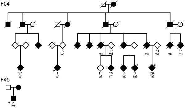
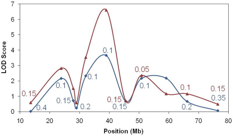
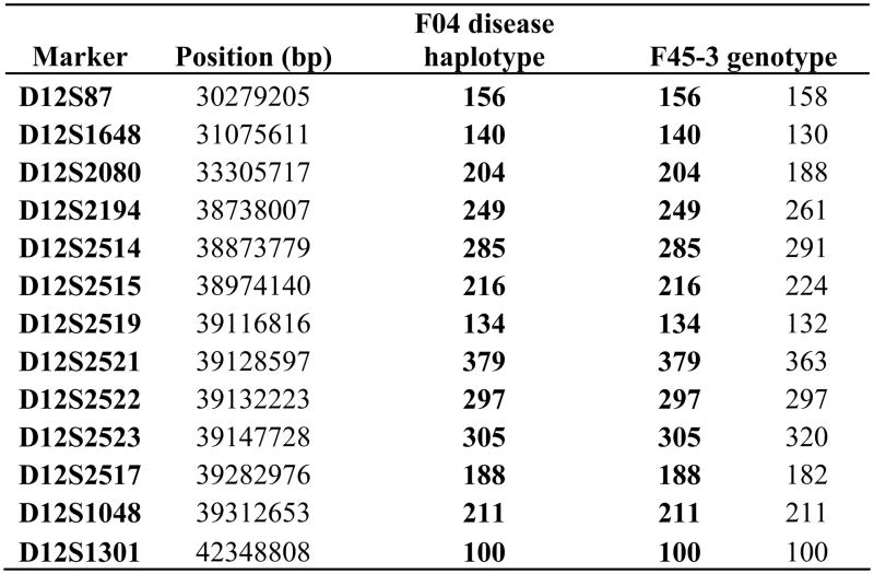
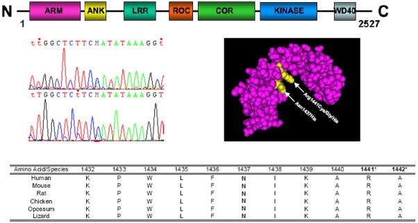
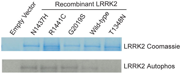
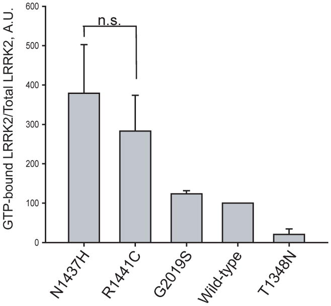
Genealogical investigation of a large Norwegian family (F04) with autosomal dominant parkinsonism has identified 18 affected family members over four generations. Genetic studies have revealed a novel pathogenic LRRK2 mutation c.4309 A>C (p.Asn1437His) that co-segregates with disease manifestation (LOD = 3.15, θ = 0). Affected carriers have an early age at onset (48 ± 7.7 SD years) and are clinically asymmetric and levodopa responsive. The variant was absent in 623 Norwegian control subjects. Further screening of patients from the same population identified one additional affected carrier (1 of 692) with familial parkinsonism who shares the same haplotype. The mutation is located within the Roc domain of the protein and enhances GTP-binding and kinase activity, further implicating these activities as the mechanisms that underlie LRRK2-linked parkinsonism.


Figures

Figure 1
Figure 1
A) Pedigrees of F04 and F45 Pedigrees of F04 and F45. Individuals with DNA available are numbered beneath symbols, the proband in each family is arrowed, males are squares, circles are females, diamonds are gender disguised. A diagonal line indicates the subject is deceased, mt = carrier, wt = non-carrier. Asterisks refer to subjects with asymmetric SPECT images. B) LOD scores Model-based, maximum two-point LOD scores are illustrated across chromosome 12. The red line shows values obtained assuming F04-1 and F04-34 are disease phenocopies, whereas the blue line was calculated assuming they have the same cause of disease. The value of θ is only given for each marker when different from 0.0. The genomic location of LRRK2 is under the peak at 39Mb. C) Disease-segregating chromosome 12q12 haplotype The higher-resolution phase-known haplotype and genotypes of F04 and F45-3 are shown.
Figure 1
Figure 1
A) Pedigrees of F04 and F45 Pedigrees of F04 and F45. Individuals with DNA available are numbered beneath symbols, the proband in each family is arrowed, males are squares, circles are females, diamonds are gender disguised. A diagonal line indicates the subject is deceased, mt = carrier, wt = non-carrier. Asterisks refer to subjects with asymmetric SPECT images. B) LOD scores Model-based, maximum two-point LOD scores are illustrated across chromosome 12. The red line shows values obtained assuming F04-1 and F04-34 are disease phenocopies, whereas the blue line was calculated assuming they have the same cause of disease. The value of θ is only given for each marker when different from 0.0. The genomic location of LRRK2 is under the peak at 39Mb. C) Disease-segregating chromosome 12q12 haplotype The higher-resolution phase-known haplotype and genotypes of F04 and F45-3 are shown.
Figure 2
Figure 2. LRRK2 idiogram of domains, chromatogram of LRRK2 c.4309 C>A, Roc domain protein structure and conservation across species
Molecular model of the Lrrk2 Roc domain highlighting in yellow the p.Asn1437His and p.Arg1441Cys/Gly/His variants was obtained from the NCBI MMDB database [MMDB ID# 64897 (PDB# 3D6T)] . Species conservation for the p.Asn1437His and adjacent amino acids was obtained from the UCSC Genome browser and the pathogenic p.Arg.1441 and putative pathogenic p.Ala1442 mutated residues are indicated with asterisks.
Figure 3
Figure 3. The p.Asn1437His mutation alters Lrrk2 GTP-binding and kinase activity
A) GTP-binding assay GTP-binding assay, where the proportion of Lrrk2 bound to GTP-γ-sepharose beads normalized to total Lrrk2 in protein lysates shows that p.Asn1437His has a similar effect on activity compared with p.Arg1441Cys mutant Lrrk2 (n.s. is non-significant) and p.Asn1437His increases the proportion of GTP-bound Lrrk2 compared with wild-type protein. Data are derived from three independent experiments. B) Lrrk2 kinase assay Lrrk2 autophosphorylation kinase assay, with total Lrrk2 indicated by coomassie stain and kinase activity indicated by autoradiography. Normalized to input protein, p.Asn1437His demonstrates enhanced kinase activity compared with wild-type protein. The results are comparable to p.Gly2019Ser and p.Arg1441Cys Lrrk2 mutant proteins. Data are representative of three experiments.

References

    1. de Lau LM, Breteler MM. Epidemiology of Parkinson’s disease. Lancet Neurol. 2006;5(6):525–535. - PubMed
    1. Farrer MJ. Genetics of Parkinson disease: paradigm shifts and future prospects. Nat Rev Genet. 2006;7(4):306–318. - PubMed
    1. Bagade S, Lill C, McQueen M, et al. The PDGene Database. Alzheimer Research Forum; 2009.
    1. Mata IF, Wedemeyer WJ, Farrer MJ, Taylor JP, Gallo KA. LRRK2 in Parkinson’s disease: protein domains and functional insights. Trends Neurosci. 2006;29(5):286–293. - PubMed
    1. West AB, Moore DJ, Choi C, et al. Parkinson’s disease-associated mutations in LRRK2 link enhanced GTP-binding and kinase activities to neuronal toxicity. Hum Mol Genet. 2007;16(2):223–232. - PubMed

Publication types

MeSH terms