. 2010 Aug;32(4):570-8.
doi: 10.1111/j.1460-9568.2010.07325.x. Epub 2010 Jul 28.

M-tropic HIV envelope protein gp120 exhibits a different neuropathological profile than T-tropic gp120 in rat striatum

Affiliations

M-tropic HIV envelope protein gp120 exhibits a different neuropathological profile than T-tropic gp120 in rat striatum

Alessia Bachis et al. Eur J Neurosci. 2010 Aug.

Abstract

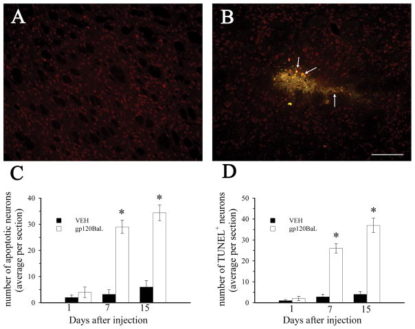
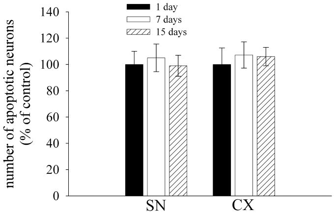
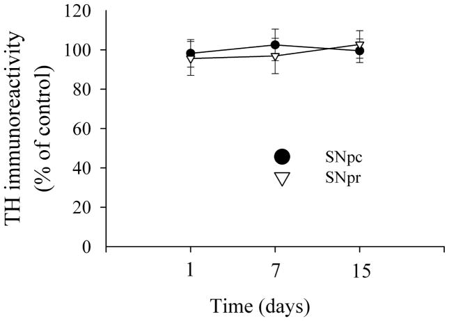
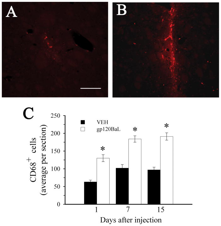
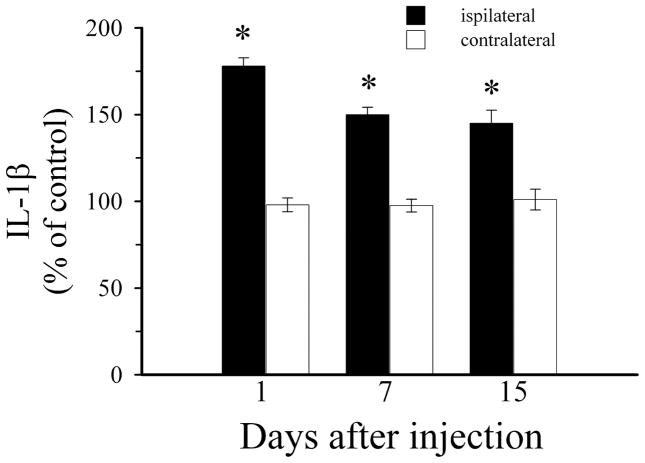
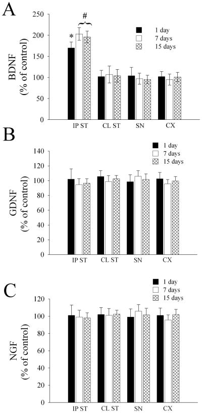
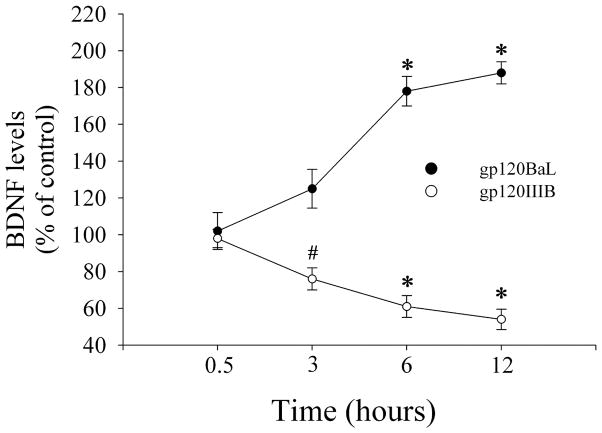
Most early human immunodeficiency virus type 1 (HIV-1) strains are macrophage (M)-tropic HIV variants and use the chemokine receptor CCR5 for infection. Neuronal loss and dementia are less severe among individuals infected with M-tropic strains. However, after several years, the T-cell (T)-tropic HIV strain, which uses the CXCR4 variant, can emerge in conjunction with brain abnormalities, suggesting strain-specific differences in neuropathogenicity. The molecular and cellular mechanisms of such diversity remain under investigation. We have previously demonstrated that HIV envelope protein gp120IIIB, which binds to CXCR4, causes neuronal apoptosis in rodents. Thus, we have used a similar experimental model to examine the neurotoxic effects of M-tropic gp120BaL. gp120BaL was microinjected in the rat striatum and neuronal apoptosis was examined in the striatum, as well as in anatomically connected areas, such as the somatosensory cortex and the substantia nigra. gp120BaL promoted neuronal apoptosis and tissue loss that were confined to the striatum. Apoptosis was associated with microglial activation and increased levels of interleukin-1beta. Intriguingly, gp120BaL increased brain-derived neurotrophic factor in the striatum. Overall, our data show that gp120BaL demonstrates a different neuropathological profile than gp120IIIB. A better understanding of the pathogenic mechanisms mediating HIV neurotoxicity is vital for developing effective neuroprotective therapies against AIDS-associated dementia complex.


Figures

Figure 1
Figure 1. gp120BaL promotes neuronal apoptosis
VEH or gp120BaL was microinjected into the rat striatum. Animals were sacrificed at the indicated times after the injection. Caspase-3, TUNEL and NeuN immunoreactivity were examined by immunohistochemistry in serial sections from the striatum. Representative sections from VEH (A) and gp120BaL (B) injected rats taken 50 μm from the injection site showing caspase-3 immunoreactivity (green) in NeuN (red) positive cells 7 days after injection. Arrows indicate examples of apoptotic neurons. Bar = 200 μm. C and D, The average number of caspase-3 and TUNEL positive neurons per section was determined by MetaMorph®. Data are expressed as the mean ± SEM of four animals per group (at least 10 sections per animal). *p<0.001 versus VEH control (1C: F = 73.663 for 7 days, F = 50.855 for 15 days; 1D: F = 89.832 for 7 days, F = 80.074).
Figure 2
Figure 2. gp120BaL does not cause apoptosis in distal areas
Rats were microinjected with VEH or gp120BaL into the striatum and were sacrificed at the indicated days after the injection. Sections from the SN and somatosensory cortex (CX) were processed for caspase-3 and NeuN. Data are the mean ± SEM of the number of caspase-3 positive neurons determined in at least 15 sections per area from five rats per group.
Figure 3
Figure 3. Analysis of TH immunoreactivity
TH immunoreactivity was analyzed in the SN of rats microinjected with VEH or gp120BaL into the striatum. TH immunoreactivity was quantified using MetaMorph® software in a series of sections from the SN pars compacta (pc) and SN pars reticulata (pr) as previously described (Nosheny et al., 2006). Data, expressed as percentage of controls, are the mean ± SEM of at least 15 sections per rat, n=5 rats per time point.
Figure 4
Figure 4. gp120BaL causes microglia activation at the site of injection
Animals were microinjected with VEH or gp120BaL into the striatum and were sacrificed at various days after the injection. Activated microglia was detected using an antibody against CD68. A and B. Representative striatal sections of VEH and gp120-treated rats, respectively, showing CD68 immunoreactivity. Sections were taken 50 μm from the injection site. Bar= 100 μm. C. CD68 positive cells were counted in the striatum at the indicated times after gp120 injection. Data, expressed as the mean ± SEM of five animals per group (10 sections per animal), are the average of the number of CD68 positive cells in an area of 0.25 mm2 per section. *p< 0.001 vs VEH (F = 37.496 for 1 day; F = 34.535 for 7 days; F = 54.983 for 15 days).
Figure 5
Figure 5. IL-1β is increased after gp120BaL
Striatal levels of IL-1βwere measured by ELISA 1, 7 and 15 days after the striatal injection of VEH or gp120BaL. Data expressed as % of VEH-treated animals are the mean ± SEM of five animals per group. *p<0.001 vs VEH (F = 166.967 for 1 day; F = 80.438 for 7 days; F = 27.695 for 15 days). The amount of IL-1βin ipsilateral VEH was 41 ± 8.2 pg/100 μg proteins.
Figure 6
Figure 6. gp120BaL increases BDNF
The levels of BDNF, GDNF and NGF were determined in the indicated brain areas 1, 7 and 15 days after gp120BaL injection into the striatum (IP ST=ipsilateral striatum, CL ST=contralateral striatum). Data are the mean ± SEM of five animals per group. *p< 0.05 vs VEH (F = 20.361); #p< 0.001 (F = 45.475 for 7 days; F = 38.738 for 15 days). BDNF, GDNF and NGF levels in the IP striatum of VEH-treated rats were (in pg/100 μg proteins): 400 ± 21; 22.5 ± 4.2; 18.5 ± 2.0, respectively.
Figure 7
Figure 7. BDNF release is differentially regulated by gp120s
Cerebellar granule cells were exposed to gp120BaL or gp120IIIB (5 nM, each) and medium was collected at the indicated time points. BDNF was determined in 100 μl fraction of the medium by ELISA. BDNF levels in the medium of control cells were 75 ± 10 pg/ml. Data are the mean ± SEM of three separate samples each group. #p<0.05 vs VEH (F = 9.914); *p<0.001 vs VEH (gp120BaL: F = 28.743 for 6 hr; F = 42.202 for 12 hr. gp120IIIB: F = 87.432 for 6 hr; F = 174.988 for 12 hr).

References

    1. Adamson DC, Wildemann B, Sasaki M, Glass JD, McArthur JC, Christov VI, Dawson TM, Dawson VL. Immunologic NO synthase: elevation in severe AIDS dementia and induction by HIV-1 gp41. Science. 1996;274:1917–1921. - PubMed
    1. Adle-Biassette H, Levy Y, Colombel M, Poron F, Natchev S, Keohane C, Gray F. Neuronal apoptosis in HIV infection in adults. Neuropathol Appl Neurobiol. 1995;21:218–227. - PubMed
    1. Ahmed F, Tessarollo L, Thiele C, Mocchetti I. Brain-derived neurotrophic factor modulates expression of chemokine receptors in the brain. Brain Res. 2008;1227:1–11. - PMC - PubMed
    1. Albright AV, Shieh JT, Itoh T, Lee B, Pleasure D, O’Connor MJ, Doms RW, Gonzalez-Scarano F. Microglia express CCR5, CXCR4, and CCR3, but of these, CCR5 is the principal coreceptor for human immunodeficiency virus type 1 dementia isolates. J Virol. 1999;73:205–213. - PMC - PubMed
    1. Altar CA, Boylan CB, Jackson C, Hershenson S, Miller J, Wiegand SJ, Lindsay RM, Hyman C. Brain-derived neurotrophic factor augments rotational behavior and nigrostriatal dopamine turnover in vivo. Proc Natl Acad Sci U S A. 1992;89:11347–11351. - PMC - PubMed

Publication types

MeSH terms