

Multifocal and metameric spinal cord arteriovenous malformations. Review of 19 cases
- PMID: 20670488
- PMCID: PMC4268662
- DOI: 10.1177/159101999900500105
Multifocal and metameric spinal cord arteriovenous malformations. Review of 19 cases
Abstract
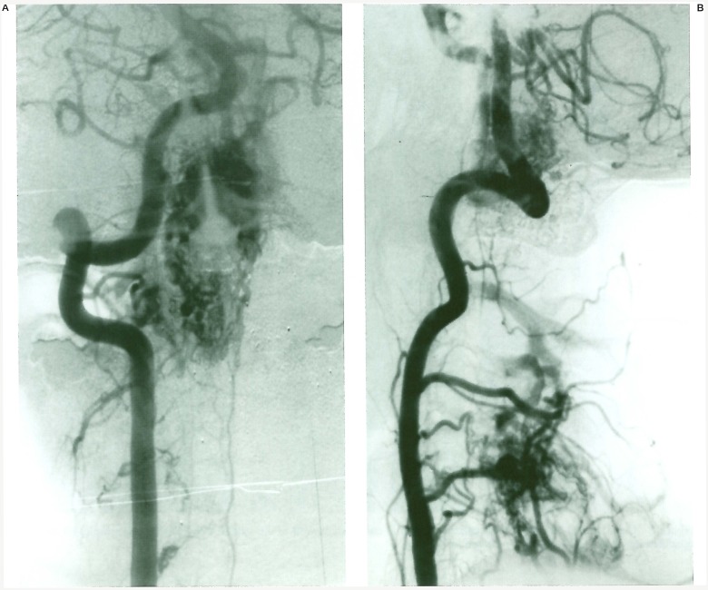
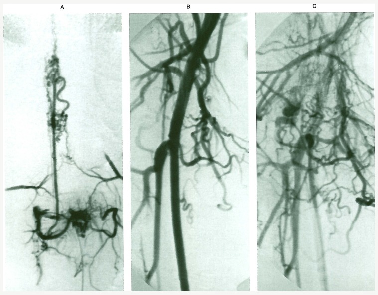
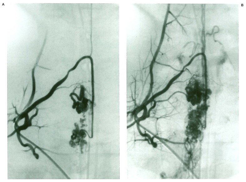
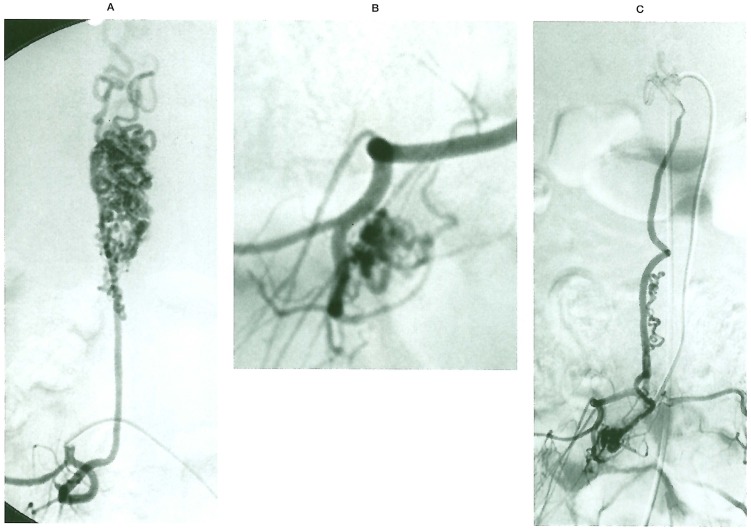
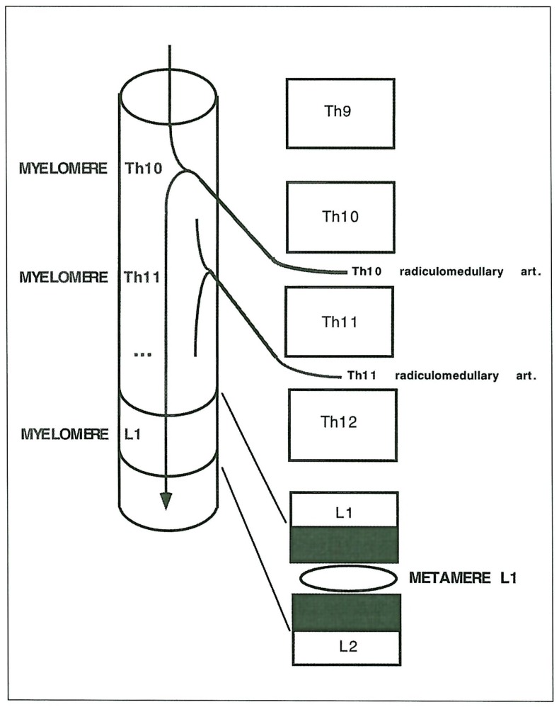
We describe 19 (16.0%) multiple vascular malformations (AVMs) in 119 spinal cord arteriovenous malformations (SCAVMs). The associated lesions were eight vertebral vascular malformations, two cutaneous, four limbs, four radicular AVMs, three bifocal SCAVMs; one patient had a bifocal cord lesion associated with vertebral and limb localisations. Various syndromic associations were seen: nine Cobb, two Klippel-Trenaunay-Weber, one Parkes Weber. An additional subgroup of unclassified associations is constituted by seven cases with bifocal intradural uni or multimetameric lesions. In our SCAVMs series, the incidence ofmultipie vascular lesions is high, in particular multifocal intradural malformations. Metameric distribution is the most frequent type of multiplicity. Identification of the myelomeric level involved in SCAVM allows segmental link between various lesions of mesodermal or neural crest origin to be discussed.
Figures
Similar articles
-
Classification of spinal cord arteriovenous shunts: proposal for a reappraisal--the Bicêtre experience with 155 consecutive patients treated between 1981 and 1999.Neurosurgery. 2002 Aug;51(2):374-9; discussion 379-80. Neurosurgery. 2002. PMID: 12182775
-
Spinal arteriovenous malformations associated with Klippel-Trenaunay-Weber syndrome: a literature search and report of two cases.AJNR Am J Neuroradiol. 2007 Mar;28(3):584-9. AJNR Am J Neuroradiol. 2007. PMID: 17353342 Free PMC article. Review.
-
Spinal cord arteriovenous malformations and the Klippel-Trenaunay-Weber syndrome.Surg Neurol. 1977 Oct;8(4):229-37. Surg Neurol. 1977. PMID: 197652
-
[Spinal arteriovenous malformations in Klippel-Trénaunay-Weber syndrome: case report].No Shinkei Geka. 1990 Sep;18(9):877-81. No Shinkei Geka. 1990. PMID: 2172852 Japanese.
-
Vascular malformations of the central nervous system: a morphological overview.Neurosurg Rev. 1986;9(3):177-216. doi: 10.1007/BF01743136. Neurosurg Rev. 1986. PMID: 3550522 Review.
Cited by
-
Pediatric metameric cervical spinal cord arteriovenous malformation managed with staged endovascular and spinal stabilization approach: a case report.Spinal Cord Ser Cases. 2024 Dec 11;10(1):80. doi: 10.1038/s41394-024-00691-w. Spinal Cord Ser Cases. 2024. PMID: 39663383
-
Craniofacial arteriovenous metameric syndrome (CAMS) 3--a transitional pattern between CAM 1 and 2 and spinal arteriovenous metameric syndromes.Neuroradiology. 2003 Sep;45(9):611-5. doi: 10.1007/s00234-003-1041-x. Epub 2003 Jul 31. Neuroradiology. 2003. PMID: 12898077
-
GNA14 and GNAQ somatic mutations cause spinal and intracranial extra-axial cavernous hemangiomas.Am J Hum Genet. 2024 Jul 11;111(7):1370-1382. doi: 10.1016/j.ajhg.2024.05.020. Epub 2024 Jun 24. Am J Hum Genet. 2024. PMID: 38917801 Free PMC article.
-
Trifocal monomyelomeric spinal cord arteriovenous fistulae in a seven-year-old boy.Interv Neuroradiol. 2001 Jun 30;7(2):121-6. doi: 10.1177/159101990100700205. Epub 2001 Jul 15. Interv Neuroradiol. 2001. PMID: 20663337 Free PMC article.
-
Cauda equina arteriovenous fistula supplied by proximal radicular artery and concomitant sacral dural arteriovenous fistula: A case report and literature review.Surg Neurol Int. 2021 Aug 16;12:405. doi: 10.25259/SNI_612_2021. eCollection 2021. Surg Neurol Int. 2021. PMID: 34513170 Free PMC article.
References
-
- Berenstein A, Lasjaunias P. Endovascular treatment of spine and spinal cord lesions. vol 5. Berlin Heidelberg New York: Springer-Verlag; 1992. Surgical Neuroangiography.
-
- Cogen P, Stein B. Spinal arteriovenous malformations with significant intramedullary components. J Neurosurg. 1983;59:471–478. - PubMed
-
- Fukushima T, Hamano K, et al. A case of multiple arteriovenous malformations and diffuse venous abnormalities with facial port-wine stain. Child's Nerv Syst. 1989;5:114–117. - PubMed
-
- Hanieh A, Blumbergs PC, et al. Arteriovenous malformations associated with soft-tissue vascular malformations: case report. J Neurosurg. 1981;54:670–672. - PubMed
-
- Iizuka Y, Rodesch G, et al. Multiple cerebral arteriovenous shunts in children: report of 13 cases. Child's Nerv Syst. 1992;8:437–444. - PubMed
LinkOut - more resources
Full Text Sources
