. 2010 Oct 27;170(3):858-64.
doi: 10.1016/j.neuroscience.2010.07.038. Epub 2010 Jul 27.

Nanostructure-initiator mass spectrometry (NIMS) imaging of brain cholesterol metabolites in Smith-Lemli-Opitz syndrome

Affiliations

Nanostructure-initiator mass spectrometry (NIMS) imaging of brain cholesterol metabolites in Smith-Lemli-Opitz syndrome

G J Patti et al. Neuroscience. .

Abstract

Cholesterol is an essential component of cellular membranes that is required for normal lipid organization and cell signaling. While the mechanisms associated with maintaining cholesterol homeostasis in the plasma and peripheral tissues have been well studied, the role and regulation of cholesterol biosynthesis in normal brain function and development have proven much more challenging to investigate. Smith-Lemli-Opitz syndrome (SLOS) is a disorder of cholesterol synthesis characterized by mutations of 7-dehydrocholesterol reductase (DHCR7) that impair the reduction of 7-dehydrocholesterol (7DHC) to cholesterol and lead to neurocognitive deficits, including cerebellar hypoplasia and austism behaviors. Here we have used a novel mass spectrometry-based imaging technique called cation-enhanced nanostructure-initiator mass spectrometry (NIMS) for the in situ detection of intact cholesterol molecules from biological tissues. We provide the first images of brain sterol localization in a mouse model for SLOS (Dhcr7(-/-)). In SLOS mice, there is a striking localization of both 7DHC and residual cholesterol in the abnormally developing cerebellum and brainstem. In contrast, the distribution of cholesterol in 1-day old healthy pups was diffuse throughout the cerebrum and comparable to that of adult mice. This study represents the first application of NIMS to localize perturbations in metabolism within pathological tissues and demonstrates that abnormal cholesterol biosynthesis may be particularly important for the development of these brain regions.


Figures

Figure 1
Figure 1
Application of NIMS to detect cholesterol metabolites dysregulated in SLOS. A, De novo synthesis of cholesterol. Cholesterol is synthesized from the precursor molecules 7DHC and 7-dehydrodemosterol. Reduction of the double bond on carbon 7 results in the formation of cholesterol and desmosterol. SLOS involves mutations of DHRC7 that impair reduction of cholesterol precursors. B, Schematic of cation-enhanced NIMS in which brain sections deposited on AgNO3-coated surfaces are desorbed/ionized by laser irradiation. A typical spectrum acquired from a 3 μm brain section is shown. Silver cationization of cholesterol results in a unique isotopic pattern separated by 2 mass units (inset).
Figure 2
Figure 2
Hematoxylin staining with corresponding cation-enhanced NIMS images of cholesterol, 7DHC, and/or desmosterol in normal and SLOS pup brains. Images are representative of those obtained from 6 different animals. A–C, Images obtained from a control littermate on postnatal day 1. Cholesterol (B) and 7DHC (C) are seen diffusely throughout the cortex. D–F, Images obtained from Dhrc−/− SLOS pups on postnatal day 1. Increased localization of cholesterol (E) and 7DHC (F) is detected in the cerebellum.
Figure 3
Figure 3
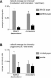
Relative quantification of cholesterol, 7DHC, and desmosterol in the developing and adult brain. (A) Ratio of average ion intensities from the developing cerebellum and brainstem of 1-day old control (n=3) and SLOS (n=3) pups with respect to the total brain. (B) Ratio of average ion intensities from the hippocampus of 1-day old control pups (n=3) and adult mice (n=3) with respect to the total brain. Data were obtained by taking the ratio of the average ion intensity over the specified brain region (see hashed circles in Figure 2B and 2E) with respect to the average ion intensity of the total brain and expressed as +/− SEM. The peak corresponding to m/z 491.26 in SLOS pups results almost exclusively from 7DHC and its average ion intensity is approximately 8 fold higher in the developing cerebellum and brainstem relative to its average ion intensity in all other brain regions.
Figure 4
Figure 4
NIMS imaging of cholesterol in embryonic mouse brain. (A) Hematoxylin and eosin staining that corresponds to the cation-enhanced NIMS image of cholesterol in a day E16–E19 embryo. The embryonic brain is indicated with a black arrow. (B) NIMS image of cholesterol in a day E16–E19 embryo. During late embryonic development the distribution of cholesterol in the brain is diffuse and does not appear to localize to any specific regions that can be resolved in the NIMS image.
Figure 5
Figure 5
Hematoxylin staining with corresponding cation-enhanced NIMS images of cholesterol, 7DHC, and/or desmosterol in adult mouse brain. Images are representative of those obtained from 3 different animals. A–C, Images obtained from a healthy, adult mouse 6–8 weeks of age. The distribution of cholesterol (B) and 7DHC (C) is diffuse throughout the brain, similar to that of healthy pups.

References

    1. Aneja A, Tierney E. Autism: the role of cholesterol in treatment. Int Rev Psychiatry. 2008;20:165–170. - PubMed
    1. Bjorkhem I, Leoni V, Meaney S. Genetic connections between neurological disorders and cholesterol metabolism. J Lipid Res. 2010 - PMC - PubMed
    1. Bjorkhem I, Meaney S. Brain cholesterol: long secret life behind a barrier. Arterioscler Thromb Vasc Biol. 2004;24:806–815. - PubMed
    1. Buitelaar JK, Willemsen-Swinkels SH. Autism: current theories regarding its pathogenesis and implications for rational pharmacotherapy. Paediatr Drugs. 2000;2:67–81. - PubMed
    1. Chugani DC, Muzik O, Rothermel R, Behen M, Chakraborty P, Mangner T, da Silva EA, Chugani HT. Altered serotonin synthesis in the dentatothalamocortical pathway in autistic boys. Ann Neurol. 1997;42:666–669. - PubMed

Publication types