. 2010 Jul 30;39(2):196-208.
doi: 10.1016/j.molcel.2010.06.018.

Defining the pathway of cytoplasmic maturation of the 60S ribosomal subunit

Affiliations

Defining the pathway of cytoplasmic maturation of the 60S ribosomal subunit

Kai-Yin Lo et al. Mol Cell. .

Abstract

In eukaryotic cells the final maturation of ribosomes occurs in the cytoplasm, where trans-acting factors are removed and critical ribosomal proteins are added for functionality. Here, we have carried out a comprehensive analysis of cytoplasmic maturation, ordering the known steps into a coherent pathway. Maturation is initiated by the ATPase Drg1. Downstream, assembly of the ribosome stalk is essential for the release of Tif6. The stalk recruits GTPases during translation. Because the GTPase Efl1, which is required for the release of Tif6, resembles the translation elongation factor eEF2, we suggest that assembly of the stalk recruits Efl1, triggering a step in 60S biogenesis that mimics aspects of translocation. Efl1 could thereby provide a mechanism to functionally check the nascent subunit. Finally, the release of Tif6 is a prerequisite for the release of the nuclear export adaptor Nmd3. Establishing this pathway provides an important conceptual framework for understanding ribosome maturation.


Figures

Figure 1
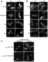
Figure 1. P0 is required for Yvh1 release
(A) The localization of NLSSV40-Yvh1-GFP (pAJ2481) was visualized in wild-type (W303) and PGAL1::P0 strain (AJY3057). Cells were diluted from galactose-containing medium into glucose-containing medium and cultured for 6 hrs. (B) Mrt4-GFP localization in wild-type (AJY3100) and PGAL1::P0 (AJY3102) strains cultured as described in A. (C) NLSSV40-Yvh1-GFP persists on 60S subunits when P0 is depleted. Strain AJY3110 (PGAL::P0 yvh1Δ) containing pAJ2481 (NLSSV40-YVH1-GFP) was cultured as described in A. Extracts were prepared and fractionated by sedimentation through 7-47% sucrose density gradients as described in (Lo et al., 2009). Western blotting was done with anti-GFP to detect NLSSV40-Yvh1-GFP and anti-Rpl8 as a marker for 60S subunits. The fraction of cells showing stronger nuclear fluorescence than cytoplasmic (fraction nuclear, fn) is given for each panel.
Figure 2
Figure 2. The assembly of the stalk is required for the function of the GTPases Efl1 and Lsg1
(A) Tif6-GFP was visualized in AJY2909 (wild-type), AJY3073 (yvh1Δ TIF6-GFP), AJY3075 (mrt4Δ TIF6-GFP), and AJY3098 (yvh1Δ mrt4Δ TIF6-GFP). (B) Nmd3(AAA)-GFP (pAJ754) was visualized in BY4741 (wild-type), AJY2976 (yvh1Δ), AJY2548 (mrt4Δ), and AJY2553 (yvh1Δ mrt4Δ). (C) Tif6-GFP was visualized in AJY3078 (wild-type) and in AJY3080 (PGAL1::P0 TIF6-GFP). Strains containing PGAL::EFL1 or PGAL::P0 were cultured in galactose or shifted to glucose medium for 24 hrs (EFL1) or 3 hrs (P0). (D) Nmd3(AAA)-GFP (pAJ754) was expressed in wild-type (W303) and PGAL1::P0 (AJY3057). (E) Pre-60S particles were immunoprecipitated from conditional Drg1, P0 and EFL1 mutants. Nmd3-myc (pAJ538) was expressed in AJY3079 (drg1-1ts TIF6-GFP), AJY3083 (PGAL1::EFL1 TIF6-GFP) and AJY3080 (PGAL1::P0 TIF6-GFP). Extracts were prepared from mid log phase cultures and immunoprecipitated as described in Materials and Methods. Proteins were separated by SDS-PAGE and western blotting was done using antibodies against c-myc (Nmd3), GFP (Tif6), Yvh1 and Rpl8.
Figure 3
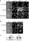
Figure 3. Depletion of P0 from HeLa cells affects the shuttling of DUSP12, MRTO4 and eIF6
(A) HeLa cells were transfected with either control siRNA or siRNA against P0 or DUSP12. The localization of MRTO4 and eIF6 was detected by indirect immunofluorescence with anti-MRTO4 (Santa Cruz Biotechnology, Inc) and anti-eIF6 antibody (Cell Signaling) 48 hours after transfection. Nuclei were visualized by staining with Hoechst 33342. (B) HeLa cells were transfected with control siRNA or siRNA against P0. After 48hr cells were transfected with DUSP12-eGFP (pcDNA3-DUSP12-EGFP). Twelve hours later cells were fixed and nuclei were stained with Hoechst 33342. (C) The efficiency of knockdown was monitored by western blotting whole cell extracts for DUSP12 and P0.
Figure 4
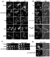
Figure 4. Mutation of sdo1 or depletion of Efl1 blocks cytoplasmic release of Nmd3 which can be suppressed by TIF6(V192F)
(A) The localization of Nmd3, Tif6 and Arx1 was examined in wild-type versus sdo1ts mutant cells. Nmd3-GFP (pAJ582), Nmd3(AAA)-GFP (pAJ754) or Tif6-GFP (pAJ1003) were expressed in wild-type (Y5563) or sdo1ts mutant (BSY28) cells. Arx1-GFP was expressed from its genomic locus in wild-type (AJY3090) and sdo1ts (AJY3086) cells. The cells were cultured at 30°C and then shifted to 37°C for 30 minutes before visualization by microscopy. (B) The localization of GFP-tagged proteins in Efl1-depleted cells. Nmd3-GFP (pAJ582) or Nmd3(AAA)-GFP (pAJ754) were expressed in wild-type (W303′) or PGAL1-EFL1 (AJY2981) cells. Tif6-GFP and Arx1-GFP were expressed from their genomic loci in wild-type (W303′) and PGAL1-EFL1 (AJY2981) cells. The cells were cultured in galactose-containing medium and then shifted to glucose medium for 26 hours before microscopy. (C) Nmd3(AAA) (pAJ754) localization was detected in PGAL1::EFL1 tif6Δ cells expressing TIF6 (AJY3013) or TIF6(V192F) (AJY3014) grown in galactose or glucose to repress EFL1 expression.
Figure 5
Figure 5. A dominant rlp24 mutant phenocopies a drg1 mutant
(A) Comparison of Rpl24 and Rlp24 generated using MACAW. Vertical bars indicate amino acid identities. The approximate truncation of the C-terminus unique to Rlp24 is indicated. (B) Serial dilutions of cultures of BY4741 containing vector, PGAL::RLP24 (pAJ2064) or PGAL::rlp24ΔC (pAJ2065) were spotted onto glucose- or galactose-containing media. (C) Extracts were prepared from wild-type (BY4741) expressing Lsg1-myc (pAJ903) or Rei1-myc (pAJ1028) in combination with Rlp24-HA (pAJ1139) or Rlp24ΔC-HA (pAJ1895) and immunoprecipitated with anti-c-myc antibody. Western blotting of SDS-PAGE separated proteins was carried out against the myc epitope, Rlp24 and Rpl8. (D) Extracts from wild-type (BY4741) expressing Rlp24-HA (pAJ1139) or Rlp24ΔC-HA (pAJ1895) in combination with either Nog1-myc (pAJ2074) or Drg1-myc (pAJ2075) were immunoprecipitated with anti-HA antibody. Western blotting was done with anti-HA or anti-myc antibodies. (E) PGAL::RLP24 (pAJ2064) and PGAL::rlp24ΔC (pAJ2065) were transformed in Arx1-GFP (AJY1948) and Tif6-GFP (AJY2909) expressing strains. Cells were grown in drop-out medium with raffinose or induced with galactose for 5 hours. (F) The localization of Arx1-GFP, Tif6-GFP, Mrt4-GFP and NLSSV40-Yvh1-GFP was visualized in W303 and drg1-1ts cells. Cells were cultured at 30°C and then shifted to 37°C for 1 hour before microscopy. (G) W303 or drg1-1ts cells expressing Lsg1-myc (pAJ903), or Rei1-myc (pAJ1028) were cultured at 30°C until OD600 ~0.5 and then shifted to 37°C for 1 hour. Immunoprecipitation was carried out with anti-c-myc antibody and protein-A beads. Precipitated proteins were eluted in Laemmli buffer and separated by SDS-PAGE. Western blotting was performed against myc, GFP (Tif6), Nmd3, Rlp24, Arx1, and Rpl8. The position of Arx1 is indicated by an arrow. The band above Arx1 that is present in all fractions is a non-specific cross-reacting protein.
Figure 6
Figure 6. Tif6 is mislocalized in both rei1Δ and jjj1Δ mutant cells and the mislocalization can be suppressed by a functional arx1 mutant
(A) The localization of Nmd3-GFP (pAJ582), Nmd3(AAA)-GFP (pAJ754) and Rlp24HA (pAJ1139) was visualized in wild-type (BY4741), rei1Δ (AJY1917) and jjj1Δ (AJY2474) cells. Tif6-GFP and Arx1-GFP were expressed genomically in the appropriate strains (see strains, Table S1). The cells were cultured at 25°C to mid-log phase. *Note that although the fraction of cells showing predominantly nuclear Tif6 is high for rei1Δ and jjj1Δ cells, in both cases, there is a uniform and significant mislocalization of Tif6 to the cytoplasm. (B) Tif6-GFP localization was visualized in wild-type (AJY2909), rei1Δ (AJY3074) and arx1Δrei1Δ (AJY3093), and in arx1Δ rei1Δ cells expressing ARX1 or arx1-S347P. (C) arx1Δ (AJY1901) or arx1Δ rei1Δ (AJY1903) cells were transformed with vector, ARX1 (pAJ2425) or arx1-S347P (pAJ1682). Ten-fold serial dilutions were plated onto selective media and incubated 3 or 4 days, for arx1Δ or arx1Δ rei1Δ, respectively. (D) The localization of Arx1-GFP (pAJ1015) or Arx1-S347P-GFP (pAJ2423) was visualized in arx1Δ rei1Δ (AJY1903) cells.
Figure 7
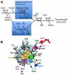
Figure 7. Proposed pathway of 60S maturation in the cytoplasm
(A) Drg1 facilitates the replacement of Rlp24 by Rpl24, which then recruits Rei1. The latter, together with Jjj1 and Ssa1/Ssa2, enables the release of the export receptor Arx1, located near the polypeptide exit tunnel. In parallel, Yvh1 enables replacement of Mrt4 with P0 to construct the ribosome stalk. In turn, the stalk recruits the GTPase Efl1 to the GTPase-associated center to release Tif6 from the subunit joining face of the particle. The release of Tif6 leads to activation of Lsg1 to release export adapter Nmd3, also from the joining face. It is important to note that the events indicated represent the order of action of these factors but not necessarily their order of association with the pre-60S particle. (B) Cartoon showing the events depicted in (A) in the context of the 60S particle in “crown” view, looking at the joining surface. Where possible, proteins have been positioned in their approximate locations on the particle. Mrt4 and Rlp24 are assumed to occupy the sites of P0 and Rpl24, respectively, in the mature subunit. The positions of Tif6 and Nmd3 are based on cryo-EM reconstructions of complexes in vitro (Gartmann et al., 2010, Sengupta et al., 2010). Arx1 is shaded to suggest that it binds on the back side of the particle, in the vicinity of the exit tunnel. CP: central protuberance; L1: L1 stalk.

References

    1. Basu U, Si K, Warner JR, Maitra U. The Saccharomyces cerevisiae TIF6 gene encoding translation initiation factor 6 is required for 60S ribosomal subunit biogenesis. Molecular and cellular biology. 2001;21:1453–1462. - PMC - PubMed
    1. Becam AM, Nasr F, Racki WJ, Zagulski M, Herbert CJ. Ria1p (Ynl163c), a protein similar to elongation factors 2, is involved in the biogenesis of the 60S subunit of the ribosome in Saccharomyces cerevisiae. Mol Genet Genomics. 2001;266:454–462. - PubMed
    1. Berk V, Cate JH. Insights into protein biosynthesis from structures of bacterial ribosomes. Current opinion in structural biology. 2007;17:302–309. - PubMed
    1. Bradatsch B, Katahira J, Kowalinski E, Bange G, Yao W, Sekimoto T, Baumgartel V, Boese G, Bassler J, Wild K, et al. Arx1 functions as an unorthodox nuclear export receptor for the 60S preribosomal subunit. Molecular cell. 2007;27:767–779. - PubMed
    1. Demoinet E, Jacquier A, Lutfalla G, Fromont-Racine M. The Hsp40 chaperone Jjj1 is required for the nucleo-cytoplasmic recycling of preribosomal factors in Saccharomyces cerevisiae. Rna. 2007;13:1570–1581. - PMC - PubMed

Publication types

MeSH terms