. 2010 Oct 8;285(41):31362-9.
doi: 10.1074/jbc.M110.150540. Epub 2010 Jul 29.

Generation of induced pluripotent stem cells in rabbits: potential experimental models for human regenerative medicine

Affiliations

Generation of induced pluripotent stem cells in rabbits: potential experimental models for human regenerative medicine

Arata Honda et al. J Biol Chem. .

Abstract

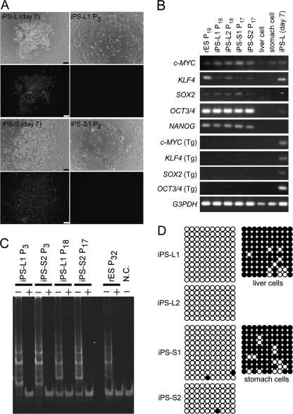
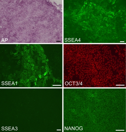
Human induced pluripotent stem (iPS) cells have the potential to establish a new field of promising regenerative medicine. Therefore, the safety and the efficiency of iPS-derived cells must be tested rigorously using appropriate animal models before human trials can commence. Here, we report the establishment of rabbit iPS cells as the first human-type iPS cells generated from a small laboratory animal species. Using lentiviral vectors, four human reprogramming genes (c-MYC, KLF4, SOX2, and OCT3/4) were introduced successfully into adult rabbit liver and stomach cells. The resulting rabbit iPS cells closely resembled human iPS cells; they formed flattened colonies with sharp edges and proliferated indefinitely in the presence of basic FGF. They expressed the endogenous pluripotency markers c-MYC, KLF4, SOX2, OCT3/4, and NANOG, whereas the introduced human genes were completely silenced. Using in vitro differentiating conditions, rabbit iPS cells readily differentiated into ectoderm, mesoderm, and endoderm. They also formed teratomas containing a variety of tissues of all three germ layers in immunodeficient mice. Thus, the rabbit iPS cells fulfilled all of the requirements for the acquisition of the fully reprogrammed state, showing high similarity to their embryonic stem cell counterparts we generated recently. However, their global gene expression analysis revealed a slight but rigid difference between these two types of rabbit pluripotent stem cells. The rabbit model should enable us to compare iPS cells and embryonic stem cells under the same standardized conditions in evaluating their ultimate feasibility for pluripotent cell-based regenerative medicine in humans.


Figures

FIGURE 1.
FIGURE 1.
Generation of iPS cells from adult rabbit liver and stomach cells. A, morphology of the primary cultures of somatic cells prepared from the liver (left panel) and stomach (right panel). B, expression of GFP at 2 days after transduction in human 4 factor-infected liver cells (left panel) and stomach cells (right panel). C, appearance of iPS cell colonies at passages 18 (liver, iPS-L1 cells) and 17 (stomach, iPS-S1 cells). D, normal number (2n = 44) of metaphase chromosomes confirmed in rabbit iPS cells arising from iPS-L1 cells at passage 18 (left panel) and iPS-S1 at passage 17 (right panel). Scale bar, 100 μm.
FIGURE 2.
FIGURE 2.
Characterization of rabbit iPS cells by their gene expression, telomerase activity, and DNA methylation of the OCT3/4 promoter. A, GFP fluorescence signal observed in the cells at 7 days after infection (iPS-L day 7 and iPS-S day 7). However, it became undetectable in iPS-L1 cells and iPS-S1 cells as early as passages 3 and 2, respectively. B, RT-PCR analysis of the expression of selected pluripotency-related genes and transgenes (Tgs) in rabbit ES cells, rabbit iPS cells, the original rabbit liver and stomach cells, and liver cells at 7 days after infection (iPS-L day 7). Five selected endogenous genes were expressed in all rabbit iPS cell lines as well as their ES cell counterparts. The four human transgenes were silenced in the iPS cells by passages 17 or 18, whereas they were slightly expressed at earlier passages (passages 6 and 7; see supplemental Fig. S2). C, detection of the telomerase activity of rabbit iPS cells using the telomeric repeat amplification protocol. The iPS cells showed high telomerase activity, similar to that of rabbit ES cells. Heat-inactivated samples (+) were used as negative controls. N.C., negative control with lysis buffer only. D, bisulfite genomic sequencing of CpG-enriched regions of the rabbit OCT3/4 promoter in iPS cells and their original (parental) somatic cells. Both liver-derived (iPS-L1 and 2, passage 7) and stomach cell-derived (iPS-S1 and 2, passage 6) lines showed a highly unmethylated pattern whereas their parental cells showed a highly methylated pattern. Open circles and filled circles represent unmethylated and methylated CpG sites, respectively. Scale bar, 100 μm.
FIGURE 3.
FIGURE 3.
Expression of pluripotency markers in rabbit iPS cells detected by staining for alkaline phosphatase (AP) activity and by immunostaining. The iPS cells were positive for alkaline phosphatase, SSEA1, SSEA4, OCT3/4, and NANOG, but not for SSEA3. Representative images were prepared from iPS-L1 cells at passage 12. Scale bars, 100 μm.
FIGURE 4.
FIGURE 4.
Differentiation of rabbit iPS cells in vitro (A) and in vivo (B). Aa, embryoid bodies formed from iPS cells under differentiation conditions at day 5. After replating on gelatin-coated dishes, they further differentiated into a variety of cell types from the three basic germ layers expressing specific markers, as detected by immunostaining. Ab, neural cells (ectoderm) expressing βIII-tubulin (green) and glial fibrillary acidic protein (red). Ac, smooth muscle cells (mesoderm) expressing α-smooth muscle myosin (green). Ad, endodermal cells expressing GATA4 (red). The cells were counterstained with DAPI. Representative images were prepared from iPS-L1 cells at passage 13. B, teratoma formation by rabbit iPS cells (iPS-L1 at passage 8). Various tissues of the three germ layer origins are identified: epidermis (a, ectoderm), bone (b, mesoderm), glands (c, endoderm), neural tissue (d, ectoderm), muscle fibers (e, mesoderm), and epithelium with goblet cells (f, endoderm). Scale bar, 100 μm.
FIGURE 5.
FIGURE 5.
DNA microarray analysis demonstrating the global gene expression profiles of iPS cells at different passage numbers, of ES cells, and of parental somatic cells. A, unsupervised hierarchical clustering. Rabbit iPS cells were more similar to each other according to their passage number rather than by origin. B, three-dimensional principal component analysis mapping. Consistent with the result of hierarchical clustering in A, iPS cells at similar passage numbers are close to each other. This map clearly demonstrates that these rabbit iPS cells started to resemble ES cells as passage proceeded. Scale bar, 100 μm.

References

    1. Martins-Taylor K., Xu R. H. (2010) J. Cell. Biochem. 109, 16–25 - PubMed
    1. Takahashi K., Yamanaka S. (2006) Cell 126, 663–676 - PubMed
    1. Takahashi K., Tanabe K., Ohnuki M., Narita M., Ichisaka T., Tomoda K., Yamanaka S. (2007) Cell 131, 861–872 - PubMed
    1. Yu J., Vodyanik M. A., Smuga-Otto K., Antosiewicz-Bourget J., Frane J. L., Tian S., Nie J., Jonsdottir G. A., Ruotti V., Stewart R., Slukvin II, Thomson J. A. (2007) Science 318, 1917–1920 - PubMed
    1. Chin M. H., Mason M. J., Xie W., Volinia S., Singer M., Peterson C., Ambartsumyan G., Aimiuwu O., Richter L., Zhang J., Khvorostov I., Ott V., Grunstein M., Lavon N., Benvenisty N., Croce C. M., Clark A. T., Baxter T., Pyle A. D., Teitell M. A., Pelegrini M., Plath K., Lowry W. E. (2009) Cell. Stem Cell 5, 111–123 - PMC - PubMed

Publication types