. 2010 Oct 8;285(41):31590-602.
doi: 10.1074/jbc.M110.101584. Epub 2010 Jul 29.

Rab32 modulates apoptosis onset and mitochondria-associated membrane (MAM) properties

Affiliations

Rab32 modulates apoptosis onset and mitochondria-associated membrane (MAM) properties

Michael Bui et al. J Biol Chem. .

Abstract

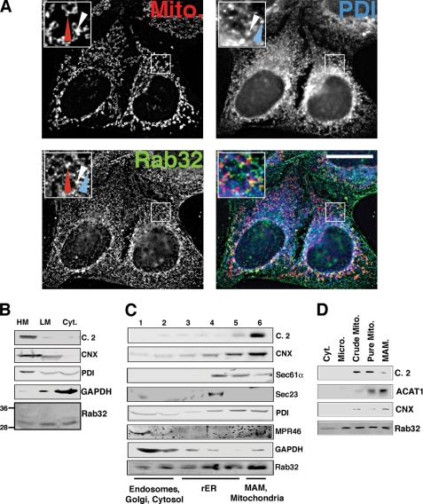
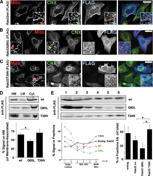
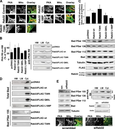
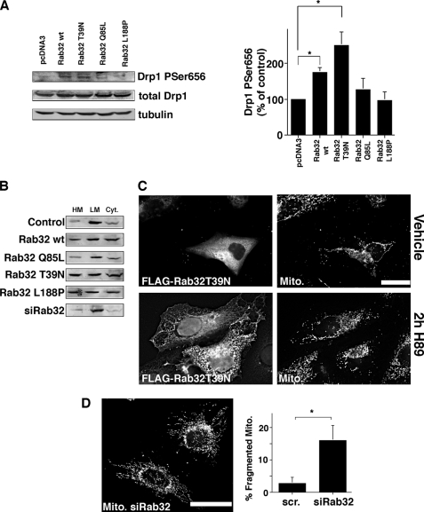
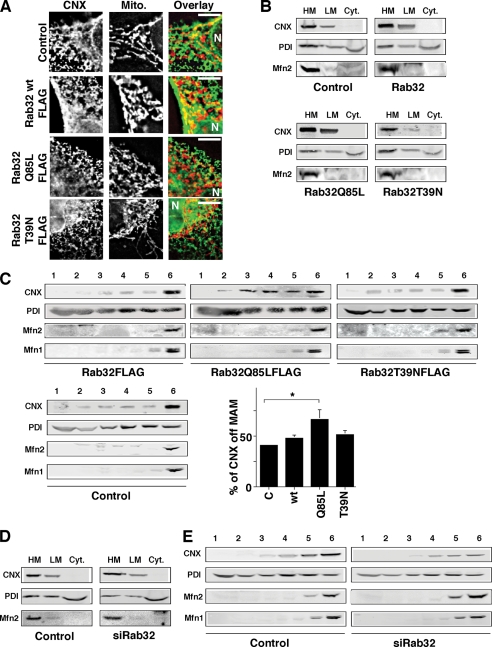
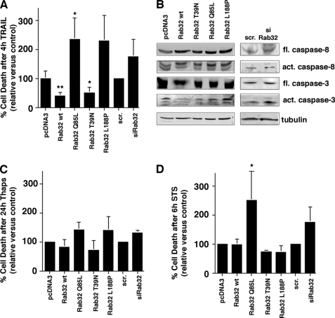
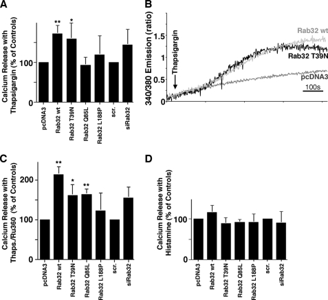
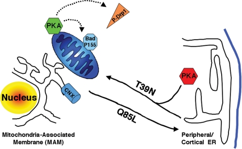
The mitochondria-associated membrane (MAM) has emerged as an endoplasmic reticulum (ER) signaling hub that accommodates ER chaperones, including the lectin calnexin. At the MAM, these chaperones control ER homeostasis but also play a role in the onset of ER stress-mediated apoptosis, likely through the modulation of ER calcium signaling. These opposing roles of MAM-localized chaperones suggest the existence of mechanisms that regulate the composition and the properties of ER membrane domains. Our results now show that the GTPase Rab32 localizes to the ER and mitochondria, and we identify this protein as a regulator of MAM properties. Consistent with such a role, Rab32 modulates ER calcium handling and disrupts the specific enrichment of calnexin on the MAM, while not affecting the ER distribution of protein-disulfide isomerase and mitofusin-2. Furthermore, Rab32 determines the targeting of PKA to mitochondrial and ER membranes and through its overexpression or inactivation increases the phosphorylation of Bad and of Drp1. Through a combination of its functions as a PKA-anchoring protein and a regulator of MAM properties, the activity and expression level of Rab32 determine the speed of apoptosis onset.


Figures

FIGURE 1.
FIGURE 1.
Intracellular localization of Rab32. A, a portion of Rab32 colocalizes with the ER. HeLa cells were grown on coverslips for 24 h and processed for immunofluorescence microscopy. Rab32 was detected with our rabbit polyclonal antibody and PDI with a mouse monoclonal antiserum (ABR, Golden, CO), and mitochondria (Mito) were preloaded with MitoTracker. Insets show a magnified area, indicated by white frames on the bigger pictures. The red arrowheads point out Rab32/mitochondria overlap, and the blue arrowheads point out Rab32/PDI overlap. An example of triple overlap is highlighted by white arrowheads. Scale bar, 25 μm. B, Rab32 fractionation into heavy (HM) and light membranes (LM) and the cytosol (Cyt). Membranes from HeLa cells were fractionated into low and high speed pellets, which were analyzed by Western blot for complex II (mitochondria), calnexin (ER/MAM), PDI (all ER), GAPDH (cytosol), and Rab32 (with molecular masses in kDa are indicated on the left). C, Rab32 distribution upon ER domain fractionation. HeLa cell homogenates were fractionated on a discontinuous 10–30% OptiPrepTM gradient. Marker proteins indicate mitochondria (complex II), MAM (calnexin), rER (Sec61α), transitional ER (Sec23), pan-ER (PDI), endosomes (MPR46), and cytosol (GAPDH). Fractions are assigned their predominant content. D, Rab32 distribution between mitochondria (Mito) and the MAM. HeLa cell homogenates were fractionated according to “Experimental Procedures.” Marker proteins indicate mitochondria (complex 2, C.2) and the MAM (acyl-CoA:cholesterol acyltransferase 1, ACAT1, and calnexin, CNX). Micro, microsomes.
FIGURE 2.
FIGURE 2.
Intracellular localization of FLAG-tagged Rab32 and its GDP/GTP-binding mutants. A, FLAG-tagged Rab32 co-localizes with the ER. HeLa cells were grown on coverslips and transfected with Rab32FLAG wild type. After 48 h, cells were processed for immunofluorescence microscopy. Rab32FLAG was detected with an anti-FLAG monoclonal antibody, calnexin (CNX), with our rabbit polyclonal antibody and mitochondria (Mito) preloaded with MitoTracker. Insets show a magnified area, indicated by white frames on the bigger pictures. Arrowheads point out Rab32/calnexin/mitochondria triple overlap. Scale bar, 25 μm. B, FLAG-tagged Rab32Q85L shows reduced overlap with mitochondria. HeLa cells were processed as in A. Arrowheads point out the absence of the Rab32/calnexin/mitochondria triple overlap. Scale bar, 25 μm. C, FLAG-tagged Rab32T39N shows overlap with mitochondria that collapse in a perinuclear area. HeLa cells were processed as in A. Arrowheads point out the Rab32/calnexin/mitochondria triple overlap. Scale bar, 25 μm. D, Rab32 GDP/GTP binding mutants show distinct fractionation patterns into heavy (HM) and light membranes (LM) and the cytosol (Cyt). Membranes from HeLa cells transfected with Rab32FLAG wild type, Q85L and T39N were fractionated into low and high speed pellets, which were analyzed by Western blot for the FLAG tag. Results from three independent fractionations were quantified. p = 0.05 between wild type and Rab32Q85L. *, p <0.01. E, Rab32 GDP/GTP binding mutants show distinct ER domain fractionation patterns. Homogenates from HeLa cells transfected with Rab32FLAG wild type (wt), Q85L, and T39N were fractionated on a discontinuous 10–30% OptiPrepTM gradient. The presence of FLAG-tagged Rab32 constructs was detected by Western blot. Results from four independent fractionations were quantified. For clarity, error bars were omitted. Additionally, the graph on the right quantifies the amounts of signal found in fractions 5 and 6 (p = 0.025 between Rab32Q85L and Rab32T39N and p = 0.025 between wild type and Rab32T39N). tER, transitional ER; Endog, endogenous.
FIGURE 3.
FIGURE 3.
Activity and the expression level of Rab32 affects the intracellular distribution of PKA and its intracellular signaling. A, overlap of PKA with mitochondria (Mito) depends on Rab32 activity. HeLa cells were grown on coverslips and transfected with an empty plasmid (pcDNA3) and pcDNA3 containing the cDNA of Rab32FLAG wild type, Rab32FLAG Q85L, Rab32FLAG T39N, and Rab32FLAG L188P. After 48 h, cells were processed for immunofluorescence microscopy, and expressing cells were identified using the FLAG signal (data not shown). PKA RII was detected with a rabbit polyclonal antibody, and mitochondria were preloaded with MitoTracker. Images show portions of cells. The position of the nucleus is indicated by the letter N. Scale bar, 10 μm. B, distribution of PKA into heavy membranes (HM), light membranes (LM), and the cytosol (Cyt) is modulated by the activity of Rab32. Membranes from HeLa control cells or cells overexpressing Rab32 and Rab32 mutants as indicated were fractionated into low and high speed pellets and the cytosol, which were analyzed by Western blot for PKA RII. C, phosphorylation of Bad on serine 155 depends on the expression level and activity of Rab32. HeLa control cells or cells overexpressing Rab32 and Rab32 mutants as indicated were lysed, and lysates were analyzed by Western blot for the presence of Bad phosphorylated on serines 112, 136, and 155, as indicated. Amounts were normalized with the signals for Bad and tubulin, and transfected Rab32 was detected using the FLAG tag of all constructs. p = 0.025 between pCDNA3 and wild type Rab32. D, distribution of Bad into heavy membranes, light membranes, and the cytosol is not affected by Rab32. Membranes from HeLa control cells or cells overexpressing Rab32 and Rab32 mutants as indicated were fractionated into low and high speed pellets and the cytosol, which were analyzed by Western blot for Bad, and Bad was phosphorylated on serine 155. Note: the information of the Bad serine 155 blots is limited to the localization of this phosphoprotein and not to levels of phosphorylation. E, Rab32 expression levels influence Bad serine 155 phosphorylation and PKA localization. Top left, analysis of Bad phosphorylation levels of HeLa cells transfected with scrambled (scr) siRNA or siRNA for Rab32, analyzed as in C. Top right, analysis of PKA membrane distribution in HeLa cells transfected with scrambled siRNA or siRNA (si) for Rab32, analyzed as in B. Rab32 expression levels are shown in HeLa cells transfected with scrambled siRNA and siRNA for Rab32. Bottom, HeLa cells were grown on coverslips and transfected with scrambled siRNA or siRNA for Rab32. After 48 h, cells were processed for immunofluorescence microscopy. PKA RII was detected with a rabbit polyclonal antibody and mitochondria with preloaded with MitoTracker. Images show portions of cells. The position of the nucleus is indicated by the letter N. Scale bar, 10 μm.
FIGURE 4.
FIGURE 4.
Activity and the expression levels of Rab32 regulate the activity of Drp1. A, phosphorylation of Drp1 on serine 656 depends on the activity of Rab32. HeLa control cells, cells overexpressing Rab32, and Rab32 mutants as indicated were lysed, and lysates were analyzed by Western blot for the presence of Drp1 phosphorylated on serine 656. Amounts were normalized with the signals for total Drp1 and actin, and Drp1 phosphorylated on serine 656 was quantified (n = 3). *, p < 0.05. B, Rab32 does not influence Drp1 targeting. Membranes from HeLa control cells, cells overexpressing Rab32 and Rab32 mutants, or cells where Rab32 has been knocked down as indicated were fractionated into low and high speed pellets and the cytosol, which were analyzed by Western blot for Drp1. HM, heavy membranes; LM, light membranes; Cyt, cytosol. C, Rab32 activity and expression levels influence mitochondrial membrane dynamics. Top row, expression of Rab32 FLAG T39N leads to the perinuclear clustering of mitochondria as described previously (34). MitoTracker-loaded HeLa cells were transfected with Rab32FLAG T39N, and transfected cells were identified by their positive FLAG signal (left). Bottom row, cells were transfected and processed as in the top row but incubated for 2 h with 10 μm H89. Scale bar, 25 μm. D, Rab32 knockdown leads to an increase in fragmented mitochondria. HeLa cells transfected with scrambled (scr) siRNA (data not shown) or siRNA for Rab32 were loaded with MitoTracker. 10 randomly selected images showing about 50 cells each were quantified for the percentage of cells with fragmented mitochondria (right). p = 0.025. Scale bar, 25 μm.
FIGURE 5.
FIGURE 5.
Active Rab32 disrupts the retention of calnexin on the MAM. A, overlap of calnexin (CNX) with mitochondria (Mito) depends on Rab32 activity. HeLa cells were grown on coverslips and transfected with an empty plasmid (pcDNA3) and pcDNA3 containing the cDNA of Rab32FLAG wild type, Rab32FLAGQ85L, Rab32FLAGT39N, and Rab32FLAGL188P. After 48 h, cells were processed for immunofluorescence microscopy, and expressing cells were identified using the FLAG signal (data not shown). Calnexin was detected with our rabbit polyclonal antibody, and mitochondria were preloaded with MitoTracker. Images show portions of cells. The position of the nucleus is indicated by the letter N. Scale bar, 10 μm. B, enrichment of calnexin on heavy membranes (HM) is disrupted by active Rab32Q85L. Membranes from HeLa control cells or cells overexpressing Rab32 and Rab32 mutants as indicated were fractionated into low and high speed pellets and the cytosol, which were analyzed by Western blot for calnexin. LM, light membranes; Cyt, cytosol. C, active Rab32Q85L disrupts calnexin MAM retention. Homogenates from HeLa cells transfected with Rab32FLAG wild type, Q85L, and T39N were fractionated on a discontinuous 10–30% OptiPrepTM gradient. The presence of calnexin was detected by Western blot. Results from three independent fractionations were quantified, and the amounts of calnexin not found in the MAM fractions 5 and 6 were graphed. p = 0.05 between control and Rab32Q85L. D, enrichment of calnexin on heavy membranes is not affected by Rab32 knockdown. Membranes were fractionated into low and high speed pellets and the cytosol and probed for calnexin as in B. E, Rab32 knockdown does not alter the distribution of calnexin on an OptiPrepTM gradient. Homogenates from HeLa cells transfected with scrambled siRNA or Rab32 siRNA were fractionated on a discontinuous 10–30% OptiPrepTM gradient. The presence of calnexin was detected by Western blot.
FIGURE 6.
FIGURE 6.
Rab32 influences TRAIL-mediated apoptosis onset. A, apoptosis onset upon TRAIL binding depends on Rab32 activity and expression levels. HeLa cells were transfected with plasmids coding for Rab32 and its GDP/GTP binding mutants and with scrambled (scr) or Rab32 siRNA. After 48 h, cells were incubated with 500 ng/ml TRAIL and subsequently analyzed for positive annexin V and propidium iodide signals. The amounts of dead cells were normalized to the vector and scrambled siRNA controls, and results from three independent experiments were graphed. **, p < 0.005 for wild type (wt); *, p < 0.01 for Rab32Q85L and Rab32T39N compared with pcDNA3. B, caspase activation upon TRAIL binding depends on Rab32 activity and expression levels. HeLa cells were transfected as in A. After 48 h, cells were incubated with 500 ng/ml TRAIL and subsequently analyzed by Western blot for caspases 3 and 8. fl, full length; act, active. C, apoptosis onset upon inhibition of kinases. HeLa cells were transfected with plasmids coding for Rab32 and its GDP/GTP-binding mutants and with scrambled or Rab32 siRNA. After 24 h, cells were incubated with 1 μm thapsigargin for 24 h and subsequently analyzed for positive annexin V and propidium iodide signals. The amounts of dead cells were normalized to the vector, and scrambled siRNA controls and results from three independent experiments were graphed. D, apoptosis onset upon staurosporine inhibition of kinases. HeLa cells were transfected with plasmids coding for Rab32 and its GDP/GTP binding mutants and with scrambled or Rab32 siRNA. After 48 h, cells were incubated with 1.2 μm staurosporine for 6 h and subsequently analyzed for positive annexin V and propidium iodide signals. The amounts of dead cells were normalized to the vector and scrambled siRNA controls, and results from three independent experiments were graphed. *, p < 0.01 for Rab32Q85L compared with pcDNA3.
FIGURE 7.
FIGURE 7.
Rab32 affects ER calcium handling. A, thapsigargin-mediated calcium release with altered Rab32 activity and expression levels. HeLa cells were transfected and processed as described under “Experimental Procedures.” Calcium was released into the FURA-2-loaded cytosol by the addition of 1 μm thapsigargin. Ratiometric signals were normalized to vector-transfected cells and quantified (n = 4). *, p < 0.05; **, p < 0.01; scr, scrambled. B, representative calcium release curves for the two constructs showing significant differences. Curves are derived from 2 × 10⁶ cells, thus corresponding to an averaged response for these cells. C, thapsigargin (Thaps)-mediated calcium release with altered Rab32 activity and expression levels in the presence of Ru360. HeLa cells were transfected and processed as in A in the presence of 10 μm Ru360 (n = 4). *, p < 0.05; **, p < 0.01. D, histamine-mediated calcium release with altered Rab32 activity and expression levels. HeLa cells were transfected and processed as described under “Experimental Procedures.” Calcium was released into the FURA-2-loaded cytosol by the addition of 200 μm histamine. Ratiometric signals were normalized to vector-transfected cells and quantified (n = 3).
FIGURE 8.
FIGURE 8.
Model for the role of Rab32 in MAM enrichment and PKA localization. Rab32 regulates the equilibrium between peripheral and perinuclear (MAM) calnexin (CNX). Active Rab32 (Q85L) extracts calnexin from the MAM and redistributes it to the cellular periphery. Rab32 also mediates the distribution of PKA between the cellular periphery, where it regulates melanosome biogenesis (43), and the perinuclear area, where it regulates apoptosis onset (this study). Substrates that are PKA-phosphorylated and dependent on Rab32 activity and expression levels include but are not limited to Bad and Drp1.

References

    1. Vance J. E. (1990) J. Biol. Chem. 265, 7248–7256 - PubMed
    1. Myhill N., Lynes E. M., Nanji J. A., Blagoveshchenskaya A. D., Fei H., Carmine Simmen K., Cooper T. J., Thomas G., Simmen T. (2008) Mol. Biol. Cell 19, 2777–2788 - PMC - PubMed
    1. Pinton P., Giorgi C., Siviero R., Zecchini E., Rizzuto R. (2008) Oncogene 27, 6407–6418 - PMC - PubMed
    1. Hayashi T., Rizzuto R., Hajnoczky G., Su T. P. (2009) Trends Cell Biol. 19, 81–88 - PMC - PubMed
    1. de Brito O. M., Scorrano L. (2008) Nature 456, 605–610 - PubMed

Publication types

MeSH terms