

Molecular analysis of non-small cell lung cancer identifies subsets with different sensitivity to insulin-like growth factor I receptor inhibition
- PMID: 20670944
- PMCID: PMC2952544
- DOI: 10.1158/1078-0432.CCR-10-0089
Molecular analysis of non-small cell lung cancer identifies subsets with different sensitivity to insulin-like growth factor I receptor inhibition
Retraction in
-
Retraction: molecular analysis of non-small cell lung cancer identifies subsets with different sensitivity to insulin-like growth factor i receptor inhibition.Clin Cancer Res. 2014 Jun 15;20(12):3358. doi: 10.1158/1078-0432.CCR-14-1118. Clin Cancer Res. 2014. PMID: 24928947 Free PMC article. No abstract available.
Abstract
Purpose: This study aimed to identify molecular determinants of sensitivity of non-small cell lung cancer (NSCLC) to anti-insulin-like growth factor receptor (IGF-IR) therapy.
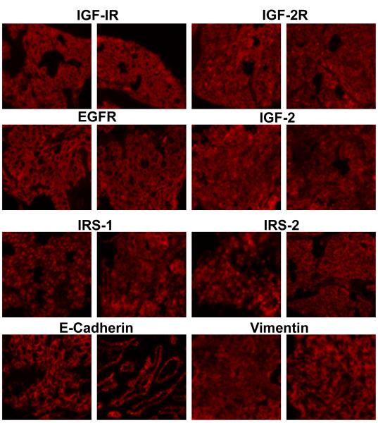
Experimental design: A total of 216 tumor samples were investigated, of which 165 consisted of retrospective analyses of banked tissue and an additional 51 were from patients enrolled in a phase II study of figitumumab, a monoclonal antibody against IGF-IR, in stage IIIb/IV NSCLC. Biomarkers assessed included IGF-IR, epidermal growth factor receptor, IGF-II, IGF-IIR, insulin receptor substrate 1 (IRS-1), IRS-2, vimentin, and E-cadherin. Subcellular localization of IRS-1 and phosphorylation levels of mitogen-activated protein kinase and Akt1 were also analyzed.
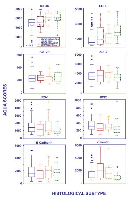
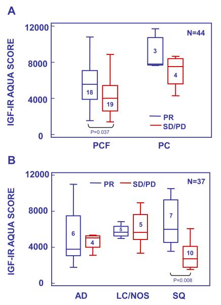
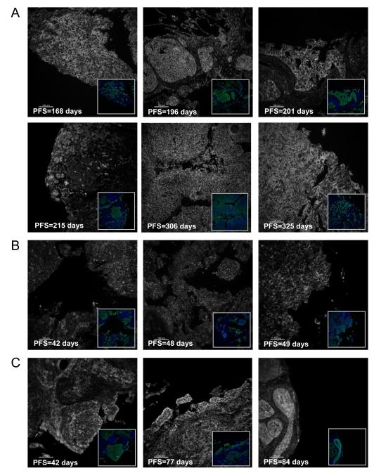
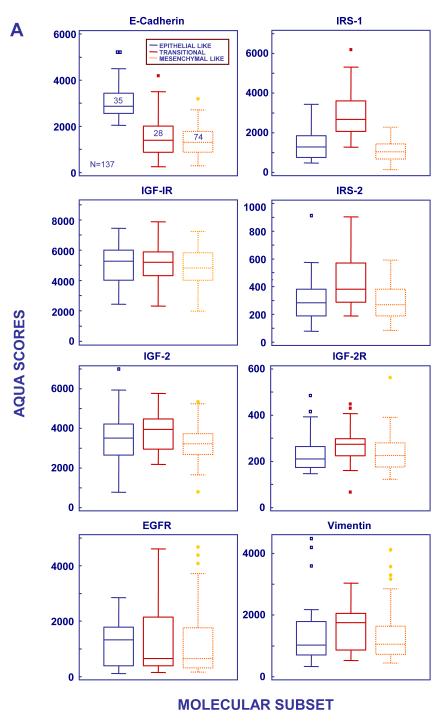
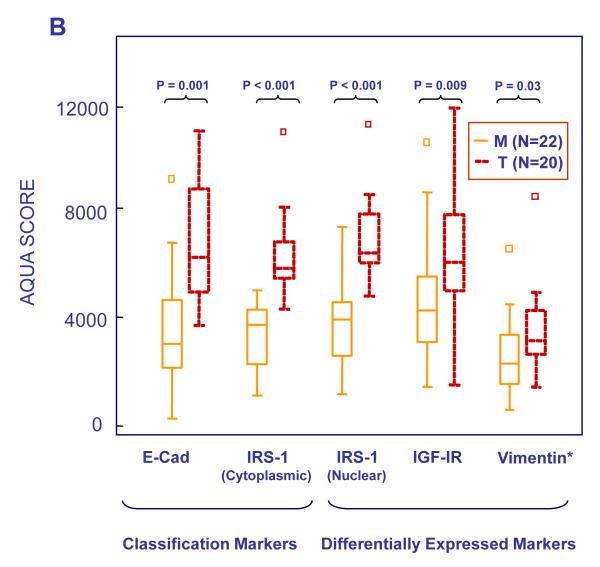
Results: IGF-IR was differentially expressed across histologic subtypes (P = 0.04), with highest levels observed in squamous cell tumors. Elevated IGF-IR expression was also observed in a small number of squamous cell tumors responding to chemotherapy combined with figitumumab (P = 0.008). Because no other biomarker/response interaction was observed using classical histologic subtyping, a molecular approach was undertaken to segment NSCLC into mechanism-based subpopulations. Principal component analysis and unsupervised Bayesian clustering identified three NSCLC subsets that resembled the steps of the epithelial to mesenchymal transition: E-cadherin high/IRS-1 low (epithelial-like), E-cadherin intermediate/IRS-1 high (transitional), and E-cadherin low/IRS-1 low (mesenchymal-like). Several markers of the IGF-IR pathway were overexpressed in the transitional subset. Furthermore, a higher response rate to the combination of chemotherapy and figitumumab was observed in transitional tumors (71%) compared with those in the mesenchymal-like subset (32%; P = 0.03). Only one epithelial-like tumor was identified in the phase II study, suggesting that advanced NSCLC has undergone significant dedifferentiation at diagnosis.
Conclusion: NSCLC comprises molecular subsets with differential sensitivity to IGF-IR inhibition.
©2010 AACR.
Figures
References
-
- Pollak M. Insulin and insulin-like growth factor signalling in neoplasia. Nat Rev Cancer. 2008;8:915–28. - PubMed
-
- Favoni RE, De Cupis A, Ravera F, et al. Expression and function of the insulin like growth factor 1 system in human non small cell lung cancer and normal lung cell lines. Int. J. Cancer. 1994;56:858–66. - PubMed
-
- Spitz MR, Barnett MJ, Goodman GE, et al. Serum Insulin like growth factor(IGF) and IGF binding protein levels and risk of lung cancer: a case control study nested in the beta carotene and retinol efficacy trial cohort. Cancer Epidemiol Biomarkers Prev. 2002;11:1413–18. - PubMed
-
- Gualberto A, Pollak M. Emerging role of insulin-like growth factor receptor inhibitors in oncology:early clinical trial results and future directions. Oncogene. 2009;28:3009–21. - PubMed
-
- Cohen BD, Baker DA, Soderstrom C, et al. Combination therapy enhances the inhibition of tumor growth with the fully human anti-type 1 insulin-like growth factor receptor monoclonal antibody figitumumab. Clin Cancer Res. 2005;11:2063–73. - PubMed
Publication types
MeSH terms
Substances
Grants and funding
LinkOut - more resources
Full Text Sources
Research Materials
Miscellaneous
