. 2010 Nov;38(21):7718-35.
doi: 10.1093/nar/gkq663. Epub 2010 Jul 29.

Genomic analysis of codon, sequence and structural conservation with selective biochemical-structure mapping reveals highly conserved and dynamic structures in rotavirus RNAs with potential cis-acting functions

Affiliations

Genomic analysis of codon, sequence and structural conservation with selective biochemical-structure mapping reveals highly conserved and dynamic structures in rotavirus RNAs with potential cis-acting functions

Wilson Li et al. Nucleic Acids Res. 2010 Nov.

Abstract

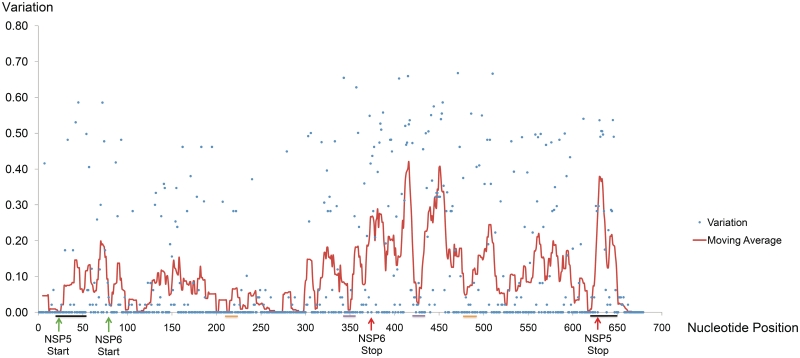
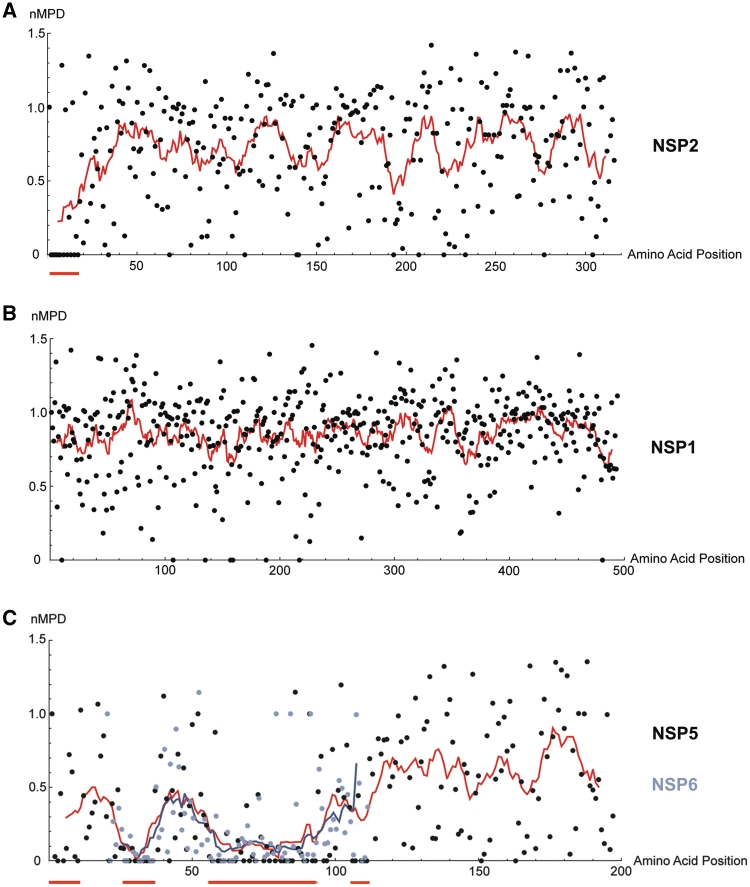
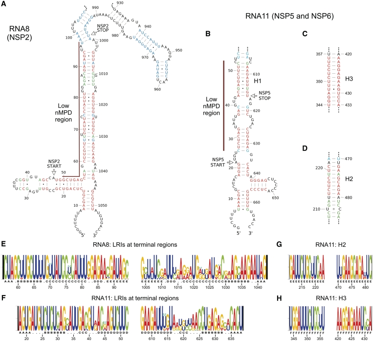
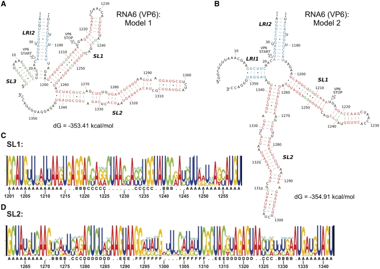
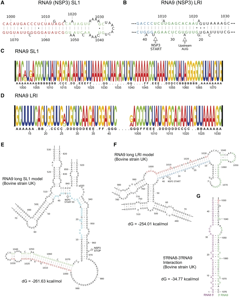
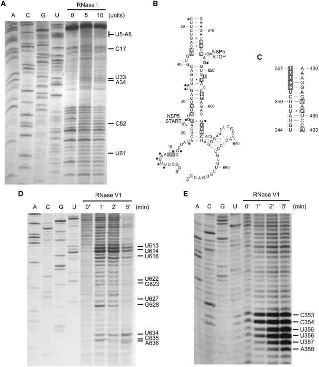
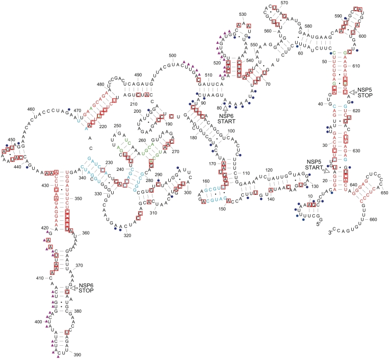
Rotaviruses are a major cause of acute, often fatal, gastroenteritis in infants and young children world-wide. Virions contain an 11 segment double-stranded RNA genome. Little is known about the cis-acting sequences and structural elements of the viral RNAs. Using a database of 1621 full-length sequences of mammalian group A rotavirus RNA segments, we evaluated the codon, sequence and RNA structural conservation of the complete genome. Codon conservation regions were found in eight ORFs, suggesting the presence of functional RNA elements. Using ConStruct and RNAz programmes, we identified conserved secondary structures in the positive-sense RNAs including long-range interactions (LRIs) at the 5' and 3' terminal regions of all segments. In RNA9, two mutually exclusive structures were observed suggesting a switch mechanism between a conserved terminal LRI and an independent 3' stem-loop structure. In RNA6, a conserved stem-loop was found in a region previously reported to have translation enhancement activity. Biochemical structural analysis of RNA11 confirmed the presence of terminal LRIs and two internal helices with high codon and sequence conservation. These extensive in silico and in vitro analyses provide evidence of the conservation, complexity, multi-functionality and dynamics of rotavirus RNA structures which likely influence RNA replication, translation and genome packaging.


Figures

Figure 1.
Figure 1.
Nucleotide variation of RNA11. Blue dots represent individual nucleotide variation values. The line graph represents moving average values using a sliding window of 9 nt (from −4 to +4 positions). Locations of start and stop codons are marked by green and red arrows, respectively. Bars indicate locations of the terminal LRIs (black), the H2 helix (orange) and the H3 helix (purple).
Figure 2.
Figure 2.
nMPD analysis of rotavirus ORFs. Results from the codon variation analysis for (A) NSP2; (B) NSP1; (C) NSP5 and NSP6 are shown. Dots represent individual nMPD scores. Line graphs show moving average values using a sliding window of 11 aa. In (C), data for NSP5 are illustrated by black dots and the red line, while data for NSP6 are illustrated by lilac dots and the purple line. Red bars underneath each plot highlight regions of high codon conservation.
Figure 3.
Figure 3.
Conserved LRIs in RNA8 and RNA11. Conserved secondary structures predicted by ConStruct and RNAz in RNA8 and RNA11 are shown, including LRIs in the terminal regions of (A) RNA8 and (B) RNA11, and two LRIs in the internal region of RNA11; (C) H3; and (D) H2. Base pairing probabilities calculated by ConStruct are represented by different colors: red (0.500 or higher), green (0.300–0.499) and blue (0.100–0.299). Regions of low-nMPD scores are marked by brown lines. Conserved structures were mapped to the sequence of the bovine rotavirus UK strain. (E and H) Nucleotide compositions of LRIs are illustrated using RNA Structure Logo for the terminal regions of RNA8 (E) and RNA11 (F), and for (G) the H2 helix and (H) the H3 helix. The height of each position indicates nucleotide conservation value. The height ratio between different bases at the same position reflects relative proportion of each base. Different letters under Structure Logos indicate the positions of individual continuous helices.
Figure 4.
Figure 4.
Stable 3′-stem–loops in RNA6. (A) Model 1 represents the consensus structure predicted using ConStruct when no constraints were applied. (B) Model 2 represents predicted structure with the 5′-terminal GGC nucleotides unpaired. Consensus structures were mapped to the bovine rotavirus UK strain sequence, and free-energy values were calculated using RNAfold. Base pairing probabilities calculated by ConStruct are represented by different colors: red (≥0.500), green (0.300–0.499) and blue (0.100–0.299). Nucleotide compositions of (C) SL1 and (D) SL2 are illustrated using RNA Structure Logo.
Figure 5.
Figure 5.
Dynamic terminal structure of RNA9. The two mutually exclusive conserved structures in RNA9 are (A) a long-stem–loop SL1 and (B) a long-stacked helix formed by LRIs. Base pairing probabilities calculated by ConStruct (A) when no constraints were applied or (B) when the UGUGACC-3′ CS was unpaired are represented by different colors: red (≥0.500), green (0.300–0.499) and blue (0.100–0.299). Nucleotide compositions of (C) SL1 and (D) long LRI are illustrated using RNA Structure Logo. (E and F) MFE structures of bovine rotavirus UK strain predicted by RNAfold using (E) SL1 and (F) long LRI as folding constraints. The 5′-strand and the 3′-strand of SL1 are highlighted in red and green, respectively. The 5′ strand of the long LRI is highlighted in blue. (G) Potential intermolecular interactions between the 5′-terminal region of RNA8 and the 3′-terminal region of RNA9 predicted by RNAcofold. Sequence from RNA8 is highlighted in purple.
Figure 6.
Figure 6.
Diverse terminal structure of RNA4. The terminal structures of three large clusters of RNA4 genotypes are represented by the MFE structure of (A) the bovine UK strain for the SA11 cluster; (B) the human ST3 strain for the Wa cluster; and (C) the human AU-1 strain for the AU-1 cluster. Base pairing probabilities calculated by ConStruct for each cluster of genotypes are represented by different colors: red (≥0.500), green (0.300–0.499) and blue (0.100–0.299). Consensus structures predicted by ConStruct using a full set of sequences with all genotypes include the 5′-terminal stem–loop and the short helix marked by the black rectangles.
Figure 7.
Figure 7.
Biochemical structure probing for RNA11. Representative auto-radiographs of primer extended products showing cleavage sites (A) by RNase I at the 5′-terminal region; (D) by RNase V1 at the 3′-terminal region; and (E) by RNase V1 close to the 5′-strand of the H3 helix. Cycle sequencing ladders are shown in the A, C, G and U lanes. Combined results from multiple experiments (B) for the terminal regions and (C) for the H3 helix are also shown, where boxes represent RNase V1 cleavages (indicating the presence of helical conformation at flanking nucleotides) and dots represent cleavages by RNases with single-strand specificities (RNases T1, U2, A, I and CL3).
Figure 8.
Figure 8.
Complete secondary structure model of RNA11. Base pairing probabilities calculated by ConStruct are represented by different colors: red (0.500 or higher), green (0.300–0.499) and blue (0.100–0.299). Red boxes represent RNase V1 cleavages (indicating the presence of helical conformation at flanking nucleotides), and blue dots represent cleavages by RNases with single-strand specificities (RNases T1, U2, A, I and CL3). Purple triangles represent potential tertiary interactions predicted by RNAz.

References

    1. Parashar UD, Gibson CJ, Bresse JS, Glass RI. Rotavirus and severe childhood diarrhea. Emerg. Infect. Dis. 2006;12:304–306. - PMC - PubMed
    1. Ruiz-Palacios GM, Perez-Schael I, Velazquez FR, Abate H, Breuer T, Clemens SC, Cheuvart B, Espinoza F, Gillard P, Innis BL, et al. Safety and efficacy of an attenuated vaccine against severe rotavirus gastroenteritis. N. Engl. J. Med. 2006;354:11–22. - PubMed
    1. Vesikari T, Matson DO, Dennehy P, Van Damme P, Santosham M, Rodriguez Z, Dallas MJ, Heyse JF, Goveia MG, Black SB, et al. Safety and efficacy of a pentavalent human-bovine (WC3) reassortant rotavirus vaccine. N. Engl. J. Med. 2006;354:23–33. - PubMed
    1. Estes MK, Kapikian AZ. In: Fields’ Virology. 5th edn. Knipe DM, Howley PM, editors. Vol. II. Philadelphia: Wolters Kluwer Health/Lippincott Williams & Wilkins; 2007. pp. 1917–1974.
    1. Cheung W, Gill M, Esposito A, Kaminski C, Courousse N, Chwetzoff S, Trugnan G, Keshavan N, Lever A, Desselberger U. Rotaviruses associate with cellular lipid droplet components to replicate in viroplasms, and compounds disrupting or blocking lipid droplets inhibit viroplasm formation and viral replication. J. Virol. 2010;84:6782–6798. - PMC - PubMed

Publication types