. 2010 Oct;154(2):820-32.
doi: 10.1104/pp.110.157560. Epub 2010 Jul 29.

Proper levels of the Arabidopsis cohesion establishment factor CTF7 are essential for embryo and megagametophyte, but not endosperm, development

Affiliations

Proper levels of the Arabidopsis cohesion establishment factor CTF7 are essential for embryo and megagametophyte, but not endosperm, development

Ling Jiang et al. Plant Physiol. 2010 Oct.

Abstract

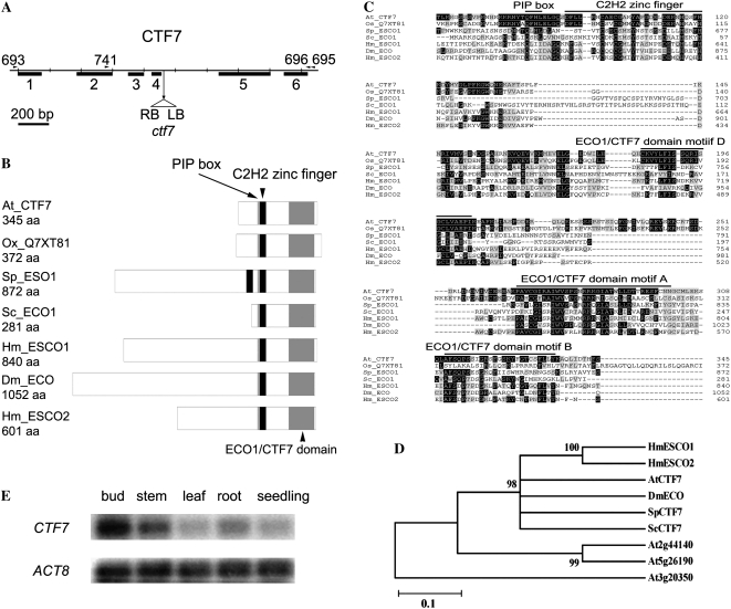
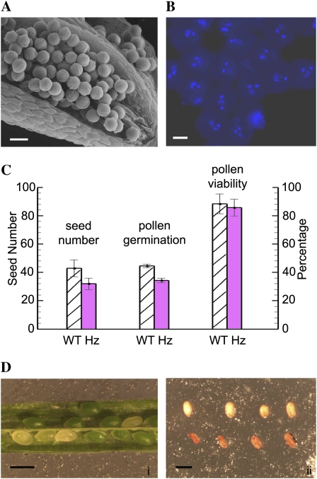
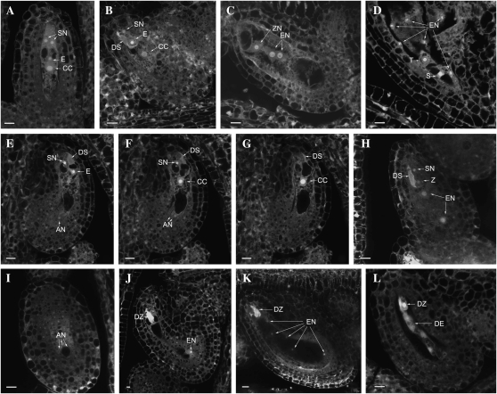
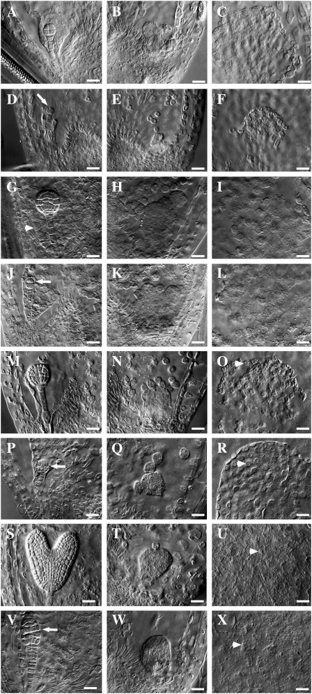
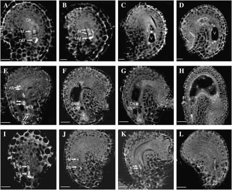
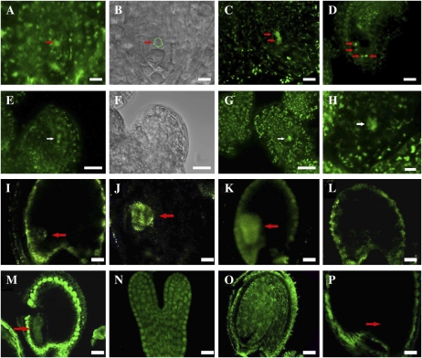
CTF7 is an essential gene in yeast that is required for the formation of sister chromatid cohesion. While recent studies have provided insights into how sister chromatid cohesion is established, less is known about how specifically CTF7 facilitates the formation of cohesion, and essentially nothing is known about how sister chromatid cohesion is established in plants. In this report, we describe the isolation and characterization of CTF7 from Arabidopsis (Arabidopsis thaliana). Arabidopsis CTF7 is similar to Saccharomyces cerevisiae CTF7 in that it lacks an amino-terminal extension, exhibits acetyltransferase activity, and can complement a yeast ctf7 temperature-sensitive mutation. CTF7 transcripts are found throughout the plant, with the highest levels present in buds. Seeds containing T-DNA insertions in CTF7 exhibit mitotic defects in the zygote. Interestingly, the endosperm developed normally in ctf7 seeds, suggesting that CTF7 is not essential for mitosis in endosperm nuclei. Minor defects were observed in female gametophytes of ctf7(+/-) plants, and plants that overexpress CTF7 exhibited female gametophyte lethality. Pollen development appeared normal in both CTF7 knockout and overexpression plants. Therefore, proper levels of CTF7 are critical for female gametophyte and embryo development but not for the establishment of mitotic cohesion during microgametogenesis or during endosperm development.


Figures

Figure 1.
Figure 1.
Arabidopsis CTF7 locus and protein structure. A, Gene map of CTF7. The exon positions and T-DNA insertion site are shown. Primers used in this study are shown above the map. LB, Left border; RB, right border. B, Schematic representations of ECO1/CTF7 proteins from different organisms. The ECO1/CTF7 domain (gray box), C2H2 zinc finger domain (black box), and PIP box (thin gray line) are shown. Accession numbers are as follows: Arabidopsis CTF7 (EU077499), O. sativa CTF7 (Q7XY81), S. pombe Eso1 (O42917), S. cerevisiae Eco1 (P43605), Homo sapiens Esco1 (Q5FWF5), Drosophila Eco (Q9VS50), H. sapiens Esco2 (Q56NI9). aa, Amino acids. C, Alignment of ECO1/CTF7 proteins in different organisms. The conserved PIP box, C2H2 zinc finger motif, and acetyltransferase domain are overlined. Identical and similar amino acids are shaded black and gray, respectively. D, Phylogenetic tree of characterized ECO1/CTF7 proteins. E, CTF7 transcripts in different tissues of Arabidopsis. ACTIN8 (ACT8) transcripts were used as a control.
Figure 2.
Figure 2.
CTF7 is the Arabidopsis cohesion establishment factor. A, Arabidopsis CTF7 exhibits acetyltransferase activity. Lanes 1 and 3, Molecular mass markers; lane 2, Coomassie Brilliant Blue-stained SDS-PAGE gel of affinity-purified CTF7-MBP; lane 4, Western blot of affinity-purified CTF7-MBP treated with anti-acetyl-Lys antibodies. Full-length CTF7-MBP is marked with arrowheads. A cleaved version of CTF7 is marked with arrows, and MBP is labeled with a star. B, Arabidopsis CTF7 can substitute for S. cerevisiae CTF7. Cultures of wild-type (WT) and temperature-sensitive ctf7-203 cells containing the pAS2 control plasmid or an Arabidopsis CTF7 complementation plasmid were grown on the appropriate selection plates at either 28°C or 33°C.
Figure 3.
Figure 3.
Pollen development in ctf7+/2 plants. A, SEM micrograph of an open ctf7+/2 stamen. B, DAPI-stained mature pollen grains from a ctf7+/2 plant. C, Histogram comparing seed number, pollen germination, and pollen viability in wild-type (WT) and ctf7+/2 plants. Data represent averages of at least 20 individuals of each genotype. D, Wild-type and ctf7 seeds: i, ctf7 seeds are normal in size but white; ii, after desiccation, ctf7 seeds are orange and shrunken (bottom row). Bars = 20 μm in A, 10 μm in B, and 1 mm in D.
Figure 4.
Figure 4.
Female gametophyte and seed development in wild-type and ctf7+/2 siliques. The images represent a single 1.5-μm optical section unless otherwise noted. A to D, Normal ovules. E to L, ctf7 ovules. A, Wild-type female gametophyte at stage FG7 containing two synergid cells (SN), an egg cell (E), and a central cell (CC). A projection of three 1.5-μm optical sections is shown. B, Female gametophyte at stage FG8 with degenerated synergid cell (DS). A projection of three 1.5-μm optical sections is shown. C, Fertilized developing seed that contains an elongated zygote (ZN) and two endosperm nuclei (EN). A projection of two 1.5-μm optical sections is shown. D, Developing seed in which the zygote has formed the suspensor (S) and terminal (T) cells. E, First optical section of an ovule with three undegenerated antipodal cells. A degenerated synergid cell, a persistent synergid cell, and the egg cell are at the micropylar pole. One of the three antipodal cells (AN) is at the bottom of the embryo sac. F, Second optical section showing two antipodal cells at the bottom of the embryo sac. G, Third optical section with the central cell in the middle of the embryo sac. H, Fertilized seed containing degenerated and persistent synergid cells and a zygote (Z). The endosperm nucleus has divided into two nuclei (EN). A projection of three 3-μm optical sections is shown. I, Optical section (3 μm) through the chalazal pole of the same seed in H. The nucleus and cell membrane of three antipodal cell nuclei are still intact. J, ctf7 seed containing a degenerated zygote (DZ) and eight endosperm nuclei. Only one of the endosperm nuclei is in the same section as the zygote. K, ctf7 seed containing a degenerated zygote and 16 endosperm nuclei. Five of the endosperm nuclei are observed. A projection of three 1.5-μm optical sections is shown. L, Degenerated ctf7 seed containing degenerated zygote and degenerated endosperm (DE) nuclei. A projection of two 1.5-μm optical sections is shown. Bars = 10 μm.
Figure 5.
Figure 5.
Embryo and endosperm development in wild-type and ctf7+/2 siliques. Wild-type (A–C, G–I, M–O, and S–U) and ctf7+/2 (D–F, J–L, P–R, and V–X) embryo and endosperm whole-mount images are shown. A, Wild-type octant-stage embryo. The MCE is still at the NCD stage. B, CZE of the embryo sac in A. C, PEN of the embryo sac in A. D, ctf7 embryo showing aberrant cell division. The arrow indicates the division plane. E, CZE of the embryo sac in D. F, PEN of the embryo sac in D. G, Wild-type globular embryo. The MCE shows signs of cellularization (arrowhead). H, CZE of the embryo sac in G. Endosperm nodules and chalazal cyst are visible. I, PEN of the embryo sac in G. J, ctf7 embryo showing aberrant cell division. The arrow indicates the division plane. K, CZE of the embryo sac in J. L, PEN of the embryo sac in J. M, Wild-type early-heart-stage embryo. N, CZE of the embryo sac in M. O, PEN of the embryo sac in M. The arrowhead indicates NCDs starting to cellularize. P, ctf7 embryo showing aberrant cell division. The arrow indicates abnormal cell shape and cell organization. Q, CZE of the embryo sac in P. R, PEN of the embryo sac in P. The arrowhead indicates NCDs starting to cellularize. S, Wild-type torpedo-stage embryo. T, CZE of the embryo sac in S. NCDs are still present. U, PEN of the embryo sac in S. The arrowhead shows cells that have completed cellularization. V, ctf7 embryo with an abnormal shape and disorganized cell order. The arrow indicates abnormal cell organization. W, CZE of the embryo sac in V. X, PEN of the embryo sac in V. The arrowhead shows cells that have completed cellularization. Bars = 10 μm.
Figure 6.
Figure 6.
Female gametophyte development revealed by laser scanning confocal microscopy in wild-type and CTF7 overexpression plants. A to H, Wild-type female gametophyte development. I to L, Female gametophyte development in CTF7 overexpression lines. A, Female gametophyte stage 1 (FG1) ovule showing the functional megaspore (M). A trace of the degraded megaspore (DM) is still visible. Ch, Chalaza. B, FG2 ovule with a two-nucleate (N) embryo sac. The degraded megaspore is still visible in some cases. C, FG3 ovule showing a late two-nucleate embryo sac with an enlarged central vacuole (V) and a small chalazal vacuole. D, Ovule with a four-nucleate embryo sac at FG4. E, Ovule with an embryo sac at FG5. Cellularization and cell differentiation are complete with the formation of two synergid nuclei (SN), an egg nucleus (EN), three antipodal nuclei (AN), and the two prominent polar nuclei (PN), which have not yet fused. F, Ovule with a mature seven-celled embryo sac at FG6. The polar nuclei have fused to form a diploid central nucleus (CN). G, Ovule at FG7 in which the antipodal cells have begun to degenerate. H, Ovule after fertilization. One synergid cell is degraded and the endosperm has completed several rounds of nuclear division. I, 35S-CTF7 ovule. No difference with wild-type FG1 ovules is apparent. The degraded megaspore is visible. J, 35S-CTF7 ovule. The embryo sac is enlarged but the female gametophyte remains at FG1. K, 35S-CTF7 ovule. The female gametophyte completed one round of mitosis. L, 35S-CTF7 ovule. The female gametophyte is degraded. Bars = 5 μm.
Figure 7.
Figure 7.
Cellular distribution of CTF7 in wild-type and 35S-CTF7::YFP plants. A to D, 35S-driven CTF7::YFP localization during female gametophyte development. A, FG1 ovule. The arrow indicates the female gametophyte nucleus. B, Bright-field image of A. The position of the female gametophyte is circled. C, FG2 ovule. The arrows indicate nuclei. D, FG7 ovule. The arrows indicate nuclei. E to H, Aborted female gametophytes in 35S-CTF7::YFP plants. E, Aborted FG1 ovule. The arrow indicates degraded nucleus. F, Bright-field image of E. G, Aborted ovule at early FG1. The arrow indicates degraded nucleus. H, Aborted ovule. The arrow indicates remnants of degraded nucleus. I, Seed with globular-stage embryo from a genomic CTF7::YFP plant. J, Seed with heart-stage embryo from a genomic CTF7::YFP transgenic plant. K, Seed with torpedo-stage embryo from a genomic CTF7::YFP transgenic plant. L, Seed from a wild-type, negative control plant. M to P, CTF7 immunolocalization on whole-mount cleared seeds. M, Torpedo-stage embryo. N, Single embryo at early torpedo stage. O, Cotyledon-stage embryo. P, Negative control with no primary antibody. Arrows in I to K, M, and P denote the position of the embryo. Bars = 5 μm.

Similar articles

Cited by

References

    1. An Y, McDowell J, Huang S, McKinney E, Chambliss S, Meagher R. (1996) Strong, constitutive expression of the Arabidopsis ACT2/ACT8 actin subclass in vegetative tissues. Plant J 10: 107–121 - PubMed
    1. Bai X, Peirsion B, Dong F, Cai X, Makaroff C. (1999) Isolation and characterization of SYN1, a RAD21-like gene essential for meiosis in Arabidopsis. Plant Cell 11: 417–430 - PMC - PubMed
    1. Bellows AM, Kenna MA, Cassimeris L, Skibbens RV. (2003) Human EFO1p exhibits acetyltransferase activity and is a unique combination of linker histone and Ctf7p/Eco1p chromatid cohesion establishment domains. Nucleic Acids Res 31: 6334–6343 - PMC - PubMed
    1. Ben-Shahar TR, Heeger S, Lehane C, East P, Flynn H, Skehel M, Uhlmann F. (2008) Eco1-dependent cohesin acetylation during establishment of sister chromatid cohesion. Science 321: 563–566 - PubMed
    1. Bhatt AM, Lister C, Page T, Fransz P, Findlay K, Jones GH, Dickinson HG, Dean C. (1999) The DIF1 gene of Arabidopsis is required for meiotic chromosome segregation and belongs to the REC8/RAD21 cohesin gene family. Plant J 19: 463–472 - PubMed

Publication types

MeSH terms

Associated data