. 2010 Jul 30;329(5991):565-8.
doi: 10.1126/science.1190181.

Early chordate origins of the vertebrate second heart field

Affiliations

Early chordate origins of the vertebrate second heart field

Alberto Stolfi et al. Science. .

Abstract

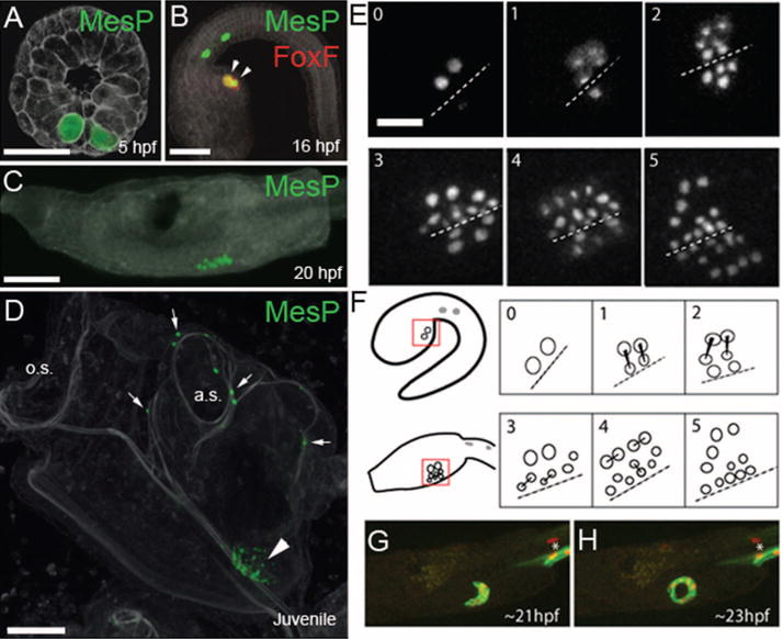
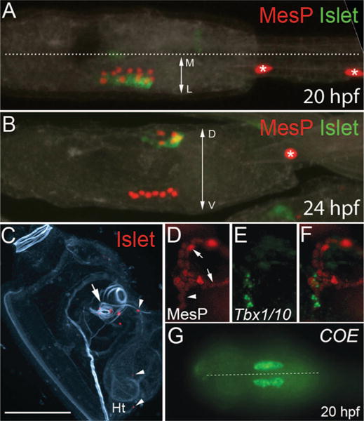
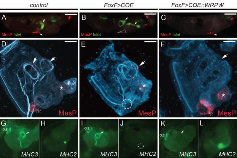
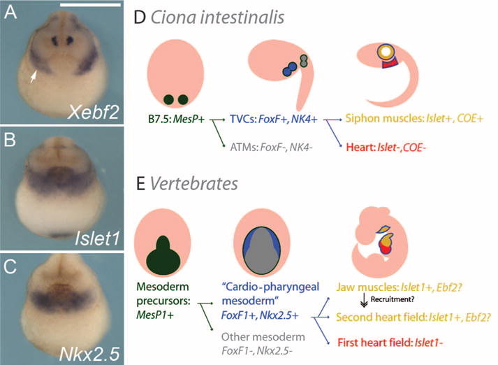
The vertebrate heart is formed from diverse embryonic territories, including the first and second heart fields. The second heart field (SHF) gives rise to the right ventricle and outflow tract, yet its evolutionary origins are unclear. We found that heart progenitor cells of the simple chordate Ciona intestinalis also generate precursors of the atrial siphon muscles (ASMs). These precursors express Islet and Tbx1/10, evocative of the splanchnic mesoderm that produces the lower jaw muscles and SHF of vertebrates. Evidence is presented that the transcription factor COE is a critical determinant of ASM fate. We propose that the last common ancestor of tunicates and vertebrates possessed multipotent cardiopharyngeal muscle precursors, and that their reallocation might have contributed to the emergence of the SHF.


Figures

Fig. 1
Fig. 1
Contribution of trunk ventral cells (TVCs) to heart and atrial siphon muscles (ASMs). (A) Immunodetection of β-gal expression (green) in B7.5 cells in a gastrulating embryo transfected with MesP>lacZ transgene. (B) Visualization of MesP>lacZ (green) and FoxF[TVC enhancer]>mCherry (red) expression in B7.5 descendants in a tailbud-stage embryo. (C) Expression of a MesP>Histone2B(H2B)∷GFP fusion protein (green) in a tadpole. (D) Visualization of MesP>H2B∷CFP (green) in a stage 38 juvenile [~100 hours post-fertilization (hpf)]. Expression is visible in the heart (arrowhead), ASMs, and longitudinal muscles (LoMs) (arrows). MesP is activated only in the B7.5 pair of cells at the gastrula stage. a.s., atrial siphon; o.s., oral siphon. (E) Frames from movie S1 (time points 0 to 2) and S2 (time points 3 to 5). Dashed line indicates ventral midline. Right-side cells are partially visible at time points 1 and 2. Stereotyped cell divisions (see text) result in four lateral TVCs on either side of the embryo flanking ~16 medial TVCs. The four lateral TVCs on either side detach and migrate to form ASMs. Medial cells form the heart. Cells were visualized as two independent time-lapse sequences of embryos transfected with MesP>H2B∷GFP/CFP. (F) Cartoon representing the events in (E). (G and H) Frames from movie S3 showing left-side ASM precursors expressing MesP>H2B∷mCherry (red) and MesP>PH∷GFP (green). ASM precursors encircle the siphon primordium, between 21 hpf (G) and 23 hpf (H). Scale bars, 50 μm [(A) to (D)], 20 μm (E). Asterisks identify anterior tail muscles (ATMs).
Fig. 2
Fig. 2
ASM-specific gene expression. (A) Dorsal view of electroporated larva exhibiting mosaic incorporation (left side) of Islet>GFP (green) and MesP>H2B∷mCherry (red) transgenes at 20 hpf, before the migration of the lateral TVCs. Islet>GFP expression is restricted to lateral TVCs. Dotted line indicates midline; M and L indicate medial and lateral, respectively. (B) Lateral view of larva expressing same transgenes as in (A) at 24 hours, after migration of Islet>GFP-positive lateral TVC descendants around the atrial siphon primordium. D, dorsal; V, ventral. (C) Juvenile (~100 hpf) raised from embryo transfected with Islet>H2B∷mCherry (red), with transgene expression visible around atrial siphons (arrow) and longitudinal muscles (arrowheads), but not heart (Ht). F-actin is stained by phallacidin (blue-green). Scale bar, 100 μm. (D) Magnified view (see fig. S7) of LoMs (arrowhead) segregating from ASMs (arrows) during metamorphosis, visualized by MesP>lacZ expression (red). Panel width is ~100 μm. (E) In situ hybridization of Tbx1/10 (green). (F) Merged view of (D) and (E), showing activation of Tbx1/10 in LoMs. (G) COE expression in lateral TVCs at 20 hpf revealed by in situ hybridization. Dotted line indicates ventral midline.
Fig. 3
Fig. 3
COE controls ASM specification. (A to C) Larvae cotransfected with MesP>H2B∷mCherry (red), Islet>GFP (green), and (A) FoxF>lacZ, (B) FoxF>COE, or (C) FoxF>COE∷WRPW. (B) All TVCs are transformed into ASM precursors (arrows). No heart primordium is formed (dashed triangle). (C) All TVC descendants form a heart (arrowhead) with no Islet>GFP expression. (D) Juveniles raised from embryos transfected with MesP>H2B∷mCherry (red), counterstained with phallacidin (blue-green). H2B∷mCherry+ B7.5 descendants populate the heart (Ht) and ASMs (arrows) (residual larval muscle staining indicated by asterisk). (E) Cotransfection with the FoxF>COE transgene, resulting in no heart (usual location indicated by dashed circle) but normal ASMs (arrow). (F) Coexpression of the FoxF>COE:WRPW transgene. TVCs form an expanded heart and there are no ASMs (arrow). (G and H) In situ hybridization on wild-type juveniles, showing MHC3 and MHC2 expression in ASMs (arrow) and heart, respectively. (I and J) FoxF>COE results in loss of MHC2 expression (dashed circle), but not MHC3 expression (arrow). (K and L) FoxF>COE∷WRPW abolishes MHC3 expression (arrow) and leads to expanded MHC2 expression. Scale bars, 50 μm; o.s., oral siphons.
Fig. 4
Fig. 4
Comparison to vertebrate pharyngeal mesoderm. (A to C) Expression of the COE ortholog Xebf2 and anterior lateral mesoderm/SHF markers Nkx2.5 and Islet1 in X. tropicalis embryos at NF stage 20. (A) Expression of Xebf2 (white arrow) partially overlaps that of Islet1 (B) and Nkx2.5 (C) in pharyngeal mesoderm lateral to the heart primordium. Scale bar, 500 μm. (D and E) The cardiopharyngeal lineages of Ciona (D) and vertebrates (E). (D) Summary of differential expression of selected regulatory genes in the B7.5 lineage. (E) Expression of orthologous genes in mouse development. Evolutionary reallocation of Islet1+ cardiopharyngeal precursors toward the heart might have given rise to the SHF.

References

    1. Buckingham M, Meilhac S, Zaffran S. Nat Rev Genet. 2005;6:826. - PubMed
    1. Abu-Issa R, Kirby ML. Annu Rev Cell Dev Biol. 2007;23:45. - PubMed
    1. Meilhac SM, Esner M, Kelly RG, Nicolas JF, Buckingham ME. Dev Cell. 2004;6:685. - PubMed
    1. Brade T, Gessert S, Kühl M, Pandur P. Dev Biol. 2007;311:297. - PubMed
    1. Gessert S, Kuhl M. Dev Biol. 2009;334:395. - PubMed

Publication types

MeSH terms

Substances