. 2010 Jul 30;329(5991):568-71.
doi: 10.1126/science.1189992.

Identification of a cell of origin for human prostate cancer

Affiliations

Identification of a cell of origin for human prostate cancer

Andrew S Goldstein et al. Science. .

Abstract

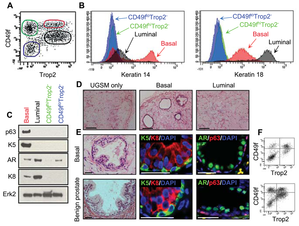
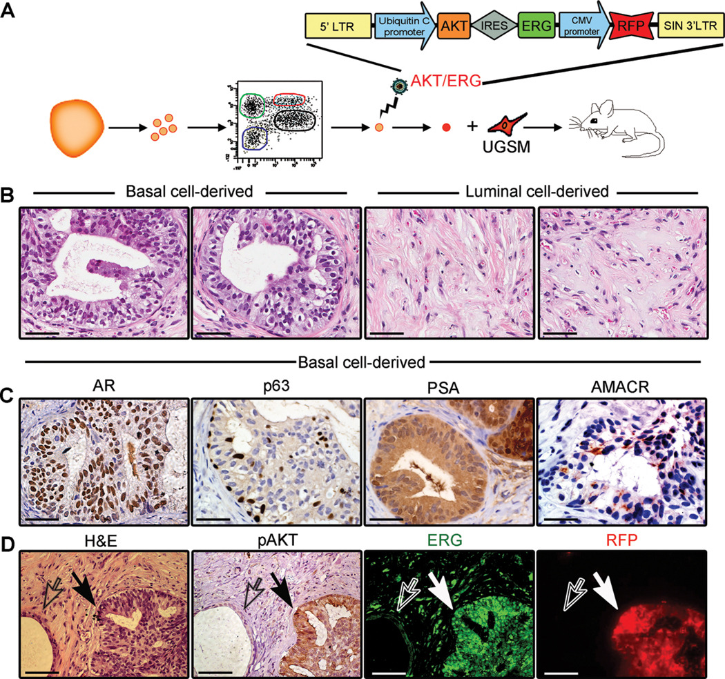
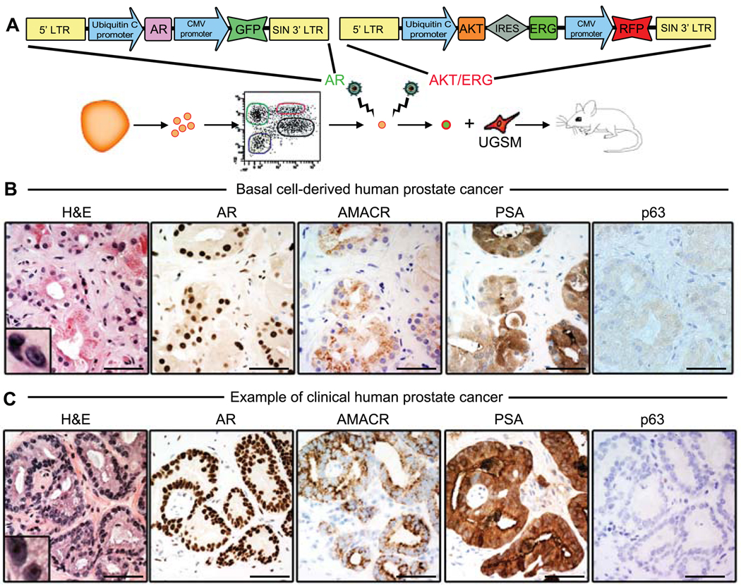
Luminal cells are believed to be the cells of origin for human prostate cancer, because the disease is characterized by luminal cell expansion and the absence of basal cells. Yet functional studies addressing the origin of human prostate cancer have not previously been reported because of a lack of relevant in vivo human models. Here we show that basal cells from primary benign human prostate tissue can initiate prostate cancer in immunodeficient mice. The cooperative effects of AKT, ERG, and androgen receptor in basal cells recapitulated the histological and molecular features of human prostate cancer, with loss of basal cells and expansion of luminal cells expressing prostate-specific antigen and alpha-methylacyl-CoA racemase. Our results demonstrate that histological characterization of cancers does not necessarily correlate with the cellular origins of the disease.


Figures

Fig. 1
Fig. 1
Purification of epithelial cell fractions from primary prostate tissue. (A) FACS plots show the distribution of dissociated primary prostate cells based on expression of CD49f and Trop2 and gates drawn to distinguish four populations. (B) Expression of Keratin 14 (K14) and Keratin 18 (K18) for each of the four populations indicated in A. (C) Immunoblots of lysates from each subpopulation analyzed for expression of basal proteins Keratin 5 (K5) and p63, and luminal proteins Keratin 8 (K8) and androgen receptor (AR). Erk2 is included as a loading control. (D) Haematoxylin and eosin (H&E) stained sections of outgrowths generated from dissociated cells transplanted subcutaneously into NSG mice. Scale bars, 200 µm. (E) Tissue generated from human prostate basal cells (top row) resembles benign human prostate tissue (bottom row). H&E shows tubule structure. High power images of immunostained prostatic tubules demonstrate presence of distinct basal and luminal layers. Yellow spots are negative for DAPI and indicate auto-fluorescent spots rather then nuclei. Scale bars, 40 µm. (F) FACS plots show presence of four populations with regards to CD49f and Trop2 staining on dissociated cells from basal-derived outgrowths or benign human prostate tissue.
Fig. 2
Fig. 2
A model of PIN initiated in primary basal cells. (A) Schematic of cell sorting, lentiviral infection (with bicistronic vector encoding activated/myristoylated AKT, ERG and the fluorescent marker RFP) and transplantation to induce initiation of PIN. (B) Images of H&E stained sections of grafts derived from transduced basal and luminal cells. Scale bars, 50 µm. (C) Immunohistochemistry of basal cell-derived lesions demonstrates prominent nuclear expression of AR with retention of p63+ cells, and cytoplasmic staining for PSA and AMACR within PIN lesions. Scale bars, 50 µm. (D) Serial sections of basal cell-derived PIN (closed arrow) next to a benign tubule (open arrow) that was not infected with lentivirus. High levels of expression of RFP (red), membrane-bound phospho-AKT (brown), and nuclear ERG (green) in PIN (closed arrow) but not in the neighboring un-infected benign tubule (open arrow). Scale bars, 100 µm.
Fig. 3
Fig. 3
A model of prostate cancer initiated in primary basal cells. (A) Schematic of cell sorting, double lentiviral infection (with GFP-encoding AR vector and bicistronic AKT/ERG vector) and transplantation to induce initiation of prostate cancer. (B–C) High-power images show similar staining patterns between basal cell-derived human prostate cancer (B) and clinical human prostate cancer (C). H&E insets demonstrate hyperchromatic nuclei with visible nucleoli at high magnification. Cancer lesions are positive for AR, AMACR and PSA and do not express the basal cell marker p63. Scale bars, 50 µm. Low power images provided in fig. S6 demonstrate heterogeneity of disease grade in both clinical human prostate cancer and basal cell-derived human prostate cancer with co-existence of benign, PIN and cancer structures.

Comment in

  • Tumorigenesis: Ground zero.
    Villanueva T. Villanueva T. Nat Rev Cancer. 2010 Sep;10(9):598. doi: 10.1038/nrc2923. Nat Rev Cancer. 2010. PMID: 20803808 No abstract available.
  • Prostate cancer from basal cells.
    Kirk R. Kirk R. Nat Rev Clin Oncol. 2010 Oct;7(10):550. doi: 10.1038/nrclinonc.2010.146. Nat Rev Clin Oncol. 2010. PMID: 20922829 No abstract available.

Similar articles

Cited by

References

    1. Pienta KJ, et al. Prostate. 2008 May 1;68:629. - PubMed
    1. Ruijter ET, van de Kaa CA, Schalken JA, Debruyne FM, Ruiter DJ. J Pathol. 1996 Nov;180:295. - PubMed
    1. Yoshimoto M, et al. Cancer Genet Cytogenet. 2006 Sep;169:128. - PubMed
    1. Tomlins SA, et al. Science. 2005 Oct 28;310:644. - PubMed
    1. Carver BS, et al. Nat Genet. 2009 May;41:619. - PMC - PubMed

Publication types

MeSH terms