. 2010 Oct 1;107(7):913-22.
doi: 10.1161/CIRCRESAHA.110.222703. Epub 2010 Jul 29.

Bone marrow mesenchymal stem cells stimulate cardiac stem cell proliferation and differentiation

Affiliations

Bone marrow mesenchymal stem cells stimulate cardiac stem cell proliferation and differentiation

Konstantinos E Hatzistergos et al. Circ Res. .

Abstract

Rationale: The regenerative potential of the heart is insufficient to fully restore functioning myocardium after injury, motivating the quest for a cell-based replacement strategy. Bone marrow-derived mesenchymal stem cells (MSCs) have the capacity for cardiac repair that appears to exceed their capacity for differentiation into cardiac myocytes.

Objective: Here, we test the hypothesis that bone marrow derived MSCs stimulate the proliferation and differentiation of endogenous cardiac stem cells (CSCs) as part of their regenerative repertoire.

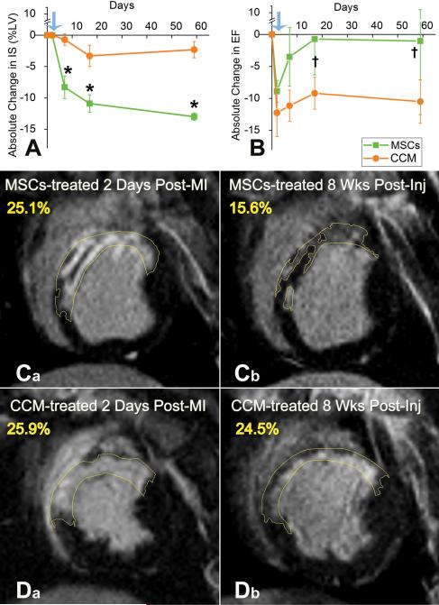
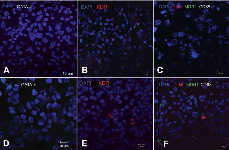
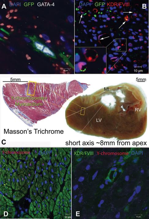
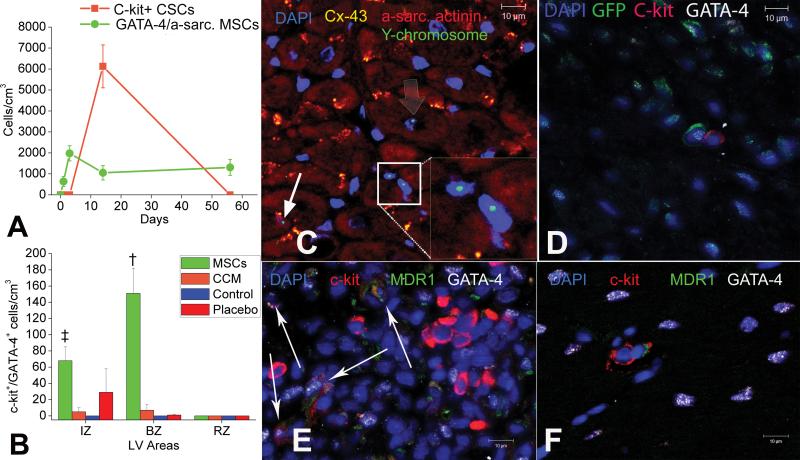
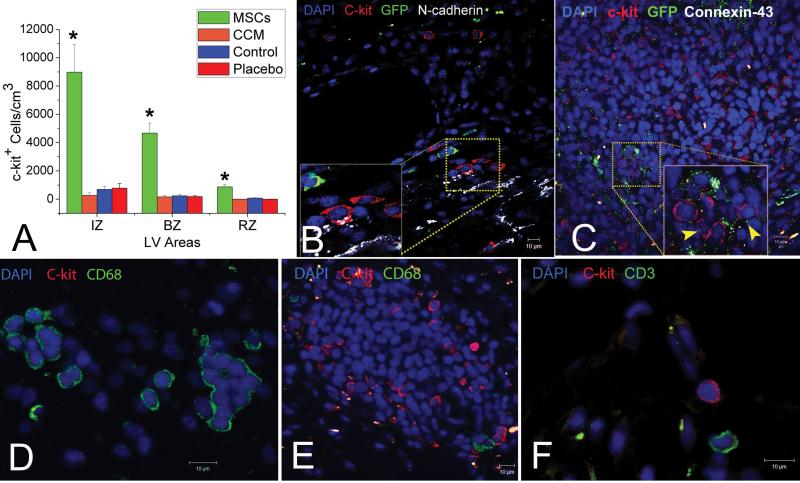
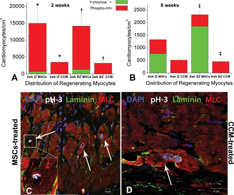
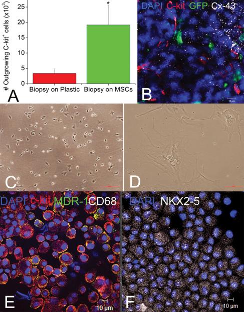
Methods and results: Female Yorkshire pigs (n=31) underwent experimental myocardial infarction (MI), and 3 days later, received transendocardial injections of allogeneic male bone marrow-derived MSCs, MSC concentrated conditioned medium (CCM), or placebo (Plasmalyte). A no-injection control group was also studied. MSCs engrafted and differentiated into cardiomyocytes and vascular structures. In addition, endogenous c-kit(+) CSCs increased 20-fold in MSC-treated animals versus controls (P<0.001), there was a 6-fold increase in GATA-4(+) CSCs in MSC versus control (P<0.001), and mitotic myocytes increased 4-fold (P=0.005). Porcine endomyocardial biopsies were harvested and plated as organotypic cultures in the presence or absence of MSC feeder layers. In vitro, MSCs stimulated c-kit(+) CSCs proliferation into enriched populations of adult cardioblasts that expressed Nkx2-5 and troponin I.

Conclusions: MSCs stimulate host CSCs, a new mechanism of action underlying successful cell-based therapeutics.


Figures

Figure 1
Figure 1. Impact of MSC and CCM on infarct size and global LV function by cardiac MRI
(A,B) Cardiac MRI documents that targeted TEI of MSCs but not CCM causes infarcted porcine hearts to improve significantly (*p=0.002), by achieving ~50% reduction in IS (A) and restoring EF (B) towards normal (p=0.042 and p=0.026 within MSCs group at 2 and 8 weeks respectively). (C,D) Representative delayed contrast hyper-enhanced images of MSCs (C a,b) and CCM (D a,b) treated animals before and 8 weeks after injections. Infarct size (yellow tracings) is reduced by MSC but not CCM administration; values in yellow correspond to scar size (%LV). Blue arrows indicate the day before TEI. Mean values± SEM (n=6 each at baseline, 4-days and 2 weeks, n=3 each at 8 weeks).
Figure 2
Figure 2. Immunophenotypic characteristics of porcine MSCs before transplantation
(A-C), Immunocytochemical staining of porcine MSCs illustrating native phenotype; all cells are negative for markers such as GATA-4 (A), KDR (B), CD68, MDR1 and c-kit (C). (D-F) Porcine Peripheral Blood Mononuclear Cells (PBMCs) used as a control cell type, for evaluating the aforementioned markers. PBMCs are negative for GATA-4 (D) but contain positive fractions for KDR (E), c-kit, CD68 and MDR1 (F).
Figure 3
Figure 3. MSCs differentiate into new cardiac myocytes and vessels
(A) A GFP+ MSC committed into cardiomyocytic lineage and undergoing symmetric division 72h after transplantation, as indicated by nuclear co-localization with GATA-4. (B) Differentiation of MSCs into endothelial lineages as indicated by co-localization of GFP with KDR and Factor-VIII related antigen (arrows). (C) Masson's trichrome stained tissue section showing the context of a Y-chromosome containing region with respect to the infarct, 2 weeks after MSCs therapy. MSCs differentiated into new myocardial tissue at the border line of a previously infarcted region. The section is located ~8mm from the apex and ~30mm from base of the LV. (D) Chimeric myocardium as indicated by the Y-chromosome containing myocytes in the cross-section of panel (C). (E) New coronary vessel formation 2 weeks after transplantation, illustrated by the Y-chromosome containing endothelial cell. [MI, myocardial infarct; LV, left ventricle; RV, right ventricle].
Figure 4
Figure 4. MSCs stimulate endogenous CSCs
(A) Graph depicting the contribution of cardiomyocyte precursors following exogenous administration of MSCs (green line) and endogenous CSCs (orange line), during cardiac repair after MI. MSC differentiation occurs rapidly after delivery. At 2-weeks, MSCs activate endogenous expansion c-kit+ CSCs (orange line). (B) Two weeks following TEI, the number of C-kit+ cells co-expressing GATA-4 is greater in MSCs vs. non-MSCs treated hearts. The cardiac precursors are preferentially located in the IZ and BZ of the MI, indicating an active process of endogenous regeneration (p=0.019 and p<0.0001) (C,D) The 2-week old chimeric myocardium contains mature cardiomyocytes (open arrow), immature MSCs (arrowheads, inset) and cardiac precursors of MSCs origin (arrow), coupled to host myocardium by connexin-43 gap junctions; Interestingly, endogenous c-kit+ CSCs are found in close proximity to MSCs (D). (E) Cluster of c-kit+ CSCs in an MSCs-treated heart; numerous CSCs are committed to cardiac lineage documented by GATA-4 and MDR-1 co-expression (arrows). (F) Few, isolated c-kit+ cells were found in non-MSC treated animals.
Figure 5
Figure 5. MSCs stimulate amplification of endogenous c-kit+ CSCs 2 weeks after injection
(A) Recruitment of c-kit+ CSCs in the MSCs-treated vs. non-treated hearts and distribution of the c-kit cells within the different zones. (*p≤0.001) (B) Endogenous c-kit+ CSCs develop putative mechanical connections with the infarcted myocardium as indicated by co-localization with N-cadherin (arrows). (C), A large cluster of c-kit+ CSCs connected to each other and to adjacent GFP+ MSCs by connexin-43. (D-F), Representative figures illustrating the non-inflammatory/mast cell phenotype of the c-kit+ CSCs. While clusters of CD68pos/c-kitneg cells could be detected in the non MSCs-treated hearts (C), the MSCs-treated hearts were rich in ckitpos/CD-68neg and CD3neg clusters of CSCs (D) Mean values ±SEM (n=6 MSCs, 3 placebo, 3 CCM and 3 Control).
Figure 6
Figure 6. MSCs stimulate endogenous cardiomyocyte cell cycling
A,B Quantification of newly formed myocytes of both host (red bargraph, phospho-H3+) and donor (green bargraph, Y-chromosome+) origin, 2 and 8 weeks post-injection respectively. MSCs stimulated host cardiomyocytes to amplify during the first 2 weeks following TEIs. The new CMs were mainly distributed at the IZ and BZ of the treated hearts, indicating active regeneration of injured myocardium. By 8 weeks endogenous cycling CMs levels had returned to normal values. Correlation between the extent of cardiomyocytes that arose from differentiated MSCs vs. amplifying host cardiomyocytes, depicted a substantially higher contribution of the latter to new cardiac muscle formation indicating the important actions of MSCs on enhancing endogenous therapeutic potentials C,D Mitotic features in endogenous cardiomyocytes from the BZ of an MSCs-treated and CCM-treated heart respectively. Mean values ±SEM (n=3 hearts, each). (* and , indicate p<0.05 between groups; indicates p=0.05 between groups).
Figure 7
Figure 7. Development of cardiac stem cell niches ex-vivo
(A) MSCs stimulate outgrowth of c-kit+ CSCs from endomyocardial biopsies. (B), Immunostaining of the primary cell cultures documents interactions between GFP+ MSCs (green) and c-kit+ cells (red) as indicated by co-localization with connexin-43 (white); these clusters resemble cardiac stem cell niches. (C, D) C-kit+ cells outgrowing after a week from the biopsy-alone are large, quiescent cells with a macrophages morphology (D); The fraction of the c-kit+ cells that were produced from the co-cultures was 3.4±0.9% of the total cell number in co-culture vs. 5.0±2.2% of the cells without co-culture. However, there were 10-fold more cells with co-culture than without, yielding many more c-kit+ cells (1.01 × 10⁶ cells/co-culture vs. 0.9 × 10⁵ cells/biopsy alone panned 1 week after plating the organotypic cultures in each group). In comparison, co-culture with MSCs egress small, semi-adherent CSCs that renew their population constantly (C). (E, F) Immunocytological stainings of c-kit+ CSCs purified and expanded from the organotypic co-cultures with MSCs, illustrate the high percentage of c-kit+ cells in MDR1 (E, yellow) and NKX2-5 (F, white), while lacking the mast-cell surface epitope CD68 (E, white). Mean values ±SEM (n=19 each).

Comment in

  • Mesenchymal stem cells express c-kit.
    Deng W. Deng W. Circ Res. 2010 Nov 12;107(10):e17; author reply e18. doi: 10.1161/CIRCRESAHA.110.230961. Circ Res. 2010. PMID: 21071709 No abstract available.

References

    1. Orlic D, Kajstura J, Chimenti S, Jakoniuk I, Anderson SM, Li B, Pickel J, McKay R, Nadal-Ginard B, Bodine DM, Leri A, Anversa P. Bone marrow cells regenerate infarcted myocardium. Nature. 2001;410:701–705. - PubMed
    1. Boyle AJ, Schulman SP, Hare JM, Oettgen P. Is stem cell therapy ready for patients? Stem Cell Therapy for Cardiac Repair. Ready for the Next Step. Circulation. 2006;114:339–352. - PubMed
    1. Hsieh PC, Segers VF, Davis ME, MacGillivray C, Gannon J, Molkentin JD, Robbins J, Lee RT. Evidence from a genetic fate-mapping study that stem cells refresh adult mammalian cardiomyocytes after injury. Nat Med. 2007;13:970–974. - PMC - PubMed
    1. Bergmann O, Bhardwaj RD, Bernard S, Zdunek S, Barnabe-Heider F, Walsh S, Zupicich J, Alkass K, Buchholz BA, Druid H, Jovinge S, Frisen J. Evidence for cardiomyocyte renewal in humans. Science. 2009;324:98–102. - PMC - PubMed
    1. Kajstura J, Urbanek K, Perl S, Hosoda T, Zheng H, Ogorek B, Ferreira-Martins J, Goichberg P, Rondon C, D'Amario D, Rota M, Del MF, Orlic D, Tisdale J, Leri A, Anversa P. Cardiomyogenesis in the Adult Human Heart. Circ Res. 2010 - PMC - PubMed

Publication types

MeSH terms

Substances