. 2010 Sep;177(3):1491-502.
doi: 10.2353/ajpath.2010.091021. Epub 2010 Jul 29.

Hypoxia increases the expression of stem-cell markers and promotes clonogenicity in glioblastoma neurospheres

Affiliations

Hypoxia increases the expression of stem-cell markers and promotes clonogenicity in glioblastoma neurospheres

Eli E Bar et al. Am J Pathol. 2010 Sep.

Abstract

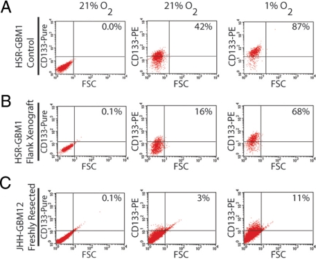
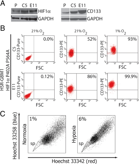
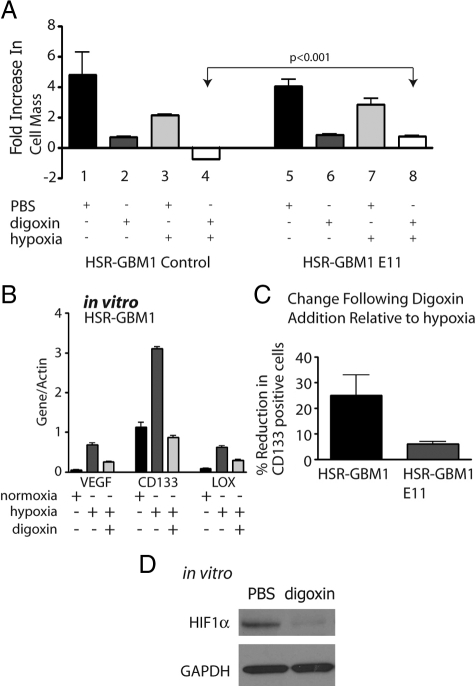
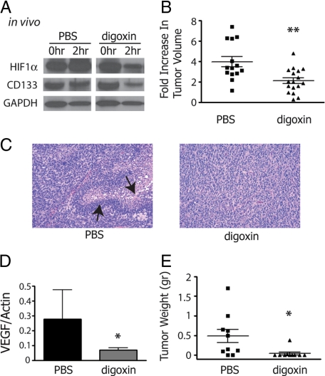
Hypoxia promotes the expansion of non-neoplastic stem and precursor cell populations in the normal brain, and is common in malignant brain tumors. We examined the effects of hypoxia on stem-like cells in glioblastoma (GBM). When GBM-derived neurosphere cultures are grown in 1% oxygen, hypoxia-inducible factor 1alpha (HIF1alpha) protein levels increase dramatically, and mRNA encoding other hypoxic response genes, such as those encoding hypoxia-inducible gene-2, lysyl oxidase, and vascular endothelial growth factor, are induced over 10-fold. Hypoxia increases the stem-like side population over fivefold, and the percentage of cells expressing CD133 threefold or more. Notch pathway ligands and targets are also induced. The rise in the stem-like fraction in GBM following hypoxia is paralleled by a twofold increase in clonogenicity. We believe HIF1alpha plays a causal role in these changes, as when oxygen-stable HIF1alpha is expressed in normoxic glioma cells CD133 is induced. We used digoxin, which has been shown to lower HIF protein levels in vitro and in vivo, to inhibit the hypoxic response. Digoxin suppressed HIF1alpha protein expression, HIF1alpha downstream targets, and slowed tumor growth in vivo. In addition, pretreatment with digoxin reduced GBM flank xenograft engraftment of hypoxic GBM cells, and daily intraperitoneal injections of digoxin were able to significantly inhibit the growth of established subcutaneous glioblastoma xenografts, and suppressed expression of vascular endothelial growth factor.


Figures

Figure 1
Figure 1
Hypoxia positively upregulates mRNA levels of lysyl oxidase (LOX), Vascular Endothelial Growth Factor (VEGF), Hypoxia Induced 2 (HIG2), and Prominin1 (CD133), and in some cells KLF4 and SOX2 in the established glioma neurosphere lines HSR-GBM2 and HSR-GBM1, and in freshly resected, GBM tumors, JHH-GBM10, JHHGBM11, and JHH-GBM12. All data was normalized to β-actin and is represented as relative expression (relative to normoxia; A). Twenty-four hours exposure to hypoxia induces HIF1α and CD133 protein expression in HSR-GBM1. GAPDH was used as a protein loading control (B). In HSR-GBM1, hypoxia positively upregulates mRNA levels of the Notch ligands, JAG1 and JAG2, as well as its targets, Hey2 and Hes1 (C). In HSR-GBM1, CD133-positive cells are two-fold more clonogenic in comparison with CD133-negative cells as indicated by a 2-fold increase in the development of large (>100mm) CSC containing spheres (**P < 0.01, two sided t-Test; D).
Figure 2
Figure 2
When the established neurosphere line HSR-GBM1 (A), xenograft isolated HSR-GBM1 (B) or freshly resected JHH-GBM12 (C) cells were exposed to hypoxia for 24 hours, the percentage of CD133-positive cells as measured by flow cytometric analysis increased 2-, 4-, and threefold, respectively. With the exception of JHH-GBM12, all experiments were repeated at least three times with similar results. Unconjugated CD133 (pure) was used as threshold control.
Figure 3
Figure 3
A: Constitutive expression of oxygen-stable HIF1αP402A/P564A (left panel) increased levels of CD133 protein expression (right panel) in HSR-GBM1 C5 and E11 as compared with the vector control infected parent line (P). B: Flow cytometric analysis for CD133 of normoxic and hypoxic HSR-GBM1 C5 and E11 cell lines overexpressing HIF1αP402A P564A showed increased baseline levels of CD133 positive cells, which further increases following 24 hours of hypoxia. Unconjugated CD133 (pure) was used as threshold control. C: Side population was increased in HSR-GBM1 cultures that were exposed for 24 hour to hypoxia as compared with normoxia grown cells.
Figure 4
Figure 4
A: Clonogenic analysis in NSMC medium. HSR-GBM2 grown under hypoxia (red bars) for 24 hours and then plated in NSMC medium showed increased clonogenic potential compared with normoxic (black bars) HSR-GBM2 cells (***P < 0.001, two sided t-test). B: HSR-GBM1 grown under hypoxia for 24 hours and then plated in NSMC medium showed increased clonogenic potential compared with normoxic HSR-GBM1control cells (P < 0.001, two sided t-test) (left panel). HSR-GBM1 stably expressing the oxygen-stable, HIF1αP402A/P564A showed maximal clonogenic potential (compared with normoxic cells from left panel) under normoxia (right panel). C: Quantification of the distribution analysis shown in B. (*P < 0.05, ns – not significant; two-sided t-test). D: Clonogenic analysis of FACS-sorted CD133 positive and negative cells showed that hypoxia affected steady-state CD133-positive cells as the average sphere diameter increases (right panel) more so than CD133-negative cells (left panel) (*P < 0.05, ***P < 0.001, two sided t-test). E: Hypoxia treatment of FACS-sorted CD133 positive and negative cells increased the number of colonies with diameter greater than 100 μm by twofold in CD133 positive but not CD133-negative cells (*P < 0.05; two-sided t-test). F: Quantitative real-time reverse transcription-PCR analysis of HIF1α and HIF2α mRNA expression in FACS sorted CD133 positive and negative cells showed that hypoxia did not affect HIF1α mRNA expression but significantly increased HIF2α expression in CD133 negative but not CD133 positive cells (***P < 0.001, ns – not significant; two-sided t-test).
Figure 5
Figure 5
In vitro digoxin treatments. A: MTT [3-(4,5-dimethylthiazol-2-yl)-diphenyl-tetrazolium bromide] analysis for HSR-GBM1 vector control and HIF1αP402A/P564A (E11) expressing cells. Growth of HSR-GBM1 was inhibited significantly in the presence of digoxin in normoxia and more so under hypoxia. Fold increase in cell mass was calculated by comparing readings on day five to those of day one. The growth of HSR-GBM1 HIF1αP402A/P564A was also inhibited by digoxin; however, in hypoxia, HIF1α appeared to completely rescue the effect digoxin under hypoxia (compare columns six and eight to columns two and four). (P < 0.001, two-sided t-test). B: Digoxin treatment reduced the expression level of the HIF1α direct targets VEGF and LOX, as well as CD133. Cells were treated with hypoxia or normoxia with PBS or digoxin (100 nmol/L) for 48 hours. C: Flow cytometric analysis of HSR-GBM1 cells, either control or E11 (Expressing oxygen stable form of HIF1α), showed that digoxin treatment reduced CD133 fraction by 25% in HSR-GBM1 control cells, an effect that was partially rescued (to 6%) by constitutive expression of the oxygen stable form of HIF1α. (The average of three independent experiments is shown. D: Western blot analysis for HIF1α in cultured HSR-GBM1 cells treated with digoxin for 24 hours. Glyceraldehyde-3-phosphate dehydrogenase used as even loading control.
Figure 6
Figure 6
In vivo digoxin treatments. A: Western blot analysis for biopsies of HSR-GBM1 flank xenografts, taken before (0 hours) and after (2 hours) intraperitoneal digoxin injection (n = 2), show reduced protein levels for CD133 and HIF1α. B: Mice treated with digoxin for 14 days showed significant reduction in tumor growth rate as compared with PBS treated mice (B, **P < 0.01 two sided t-test from 14 and 17 xenografts taken from PBS- and digoxin-treated mice, respectively). C: H&E staining of HSR-GBM1 flank xenografts treated with PBS exhibit numerous sites of palisading necrosis, which are completely absent in xenografts from mice that were treated with digoxin (arrows point to these features). D: Real-time PCR analysis for VEGF, a HIF1α dependent target gene, showed a significant reduction following digoxin treatment (*P < 0.05, two-sided t-test for pulled values from 5 PBS-treated and 6 digoxin-treated mice). E: In vitro digoxin pretreatment significantly inhibited tumor engraftment of hypoxic HSR-GBM1 cells in nude mice (*P < 0.05, two-sided t-test for pulled values from 5 PBS-treated and 5 digoxin-treated mice).

References

    1. Hegi ME, Diserens AC, Gorlia T, Hamou MF, de Tribolet N, Weller M, Kros JM, Hainfellner JA, Mason W, Mariani L, Bromberg JE, Hau P, Mirimanoff RO, Cairncross JG, Janzer RC, Stupp R. MGMT gene silencing and benefit from temozolomide in glioblastoma. N Engl J Med. 2005;352:997–1003. - PubMed
    1. Stupp R, Mason WP, van den Bent MJ, Weller M, Fisher B, Taphoorn MJ, Belanger K, Brandes AA, Marosi C, Bogdahn U, Curschmann J, Janzer RC, Ludwin SK, Gorlia T, Allgeier A, Lacombe D, Cairncross JG, Eisenhauer E, Mirimanoff RO. Radiotherapy plus concomitant and adjuvant temozolomide for glioblastoma. N Engl J Med. 2005;352:987–996. - PubMed
    1. Galli R, Binda E, Orfanelli U, Cipelletti B, Gritti A, De Vitis S, Fiocco R, Foroni C, Dimeco F, Vescovi A. Isolation and characterization of tumorigenic, stem-like neural precursors from human glioblastoma. Cancer Res. 2004;64:7011–7021. - PubMed
    1. Hemmati HD, Nakano I, Lazareff JA, Masterman-Smith M, Geschwind DH, Bronner-Fraser M, Kornblum HI. Cancerous stem cells can arise from pediatric brain tumors. Proc Natl Acad Sci USA. 2003;100:15178–15183. - PMC - PubMed
    1. Singh SK, Hawkins C, Clarke ID, Squire JA, Bayani J, Hide T, Henkelman RM, Cusimano MD, Dirks PB. Identification of human brain tumour initiating cells. Nature. 2004;432:396–401. - PubMed

Publication types

MeSH terms

Substances