Review
. 2010:2010:956071.
doi: 10.1155/2010/956071. Epub 2010 Jul 8.

The structure and function of serum opacity factor: a unique streptococcal virulence determinant that targets high-density lipoproteins

Affiliations
Review

The structure and function of serum opacity factor: a unique streptococcal virulence determinant that targets high-density lipoproteins

Harry S Courtney et al. J Biomed Biotechnol. 2010.

Abstract

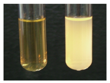
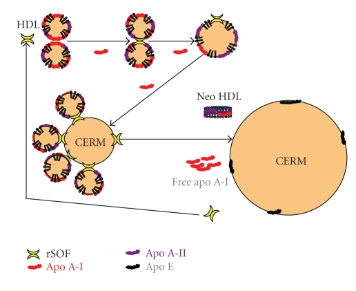
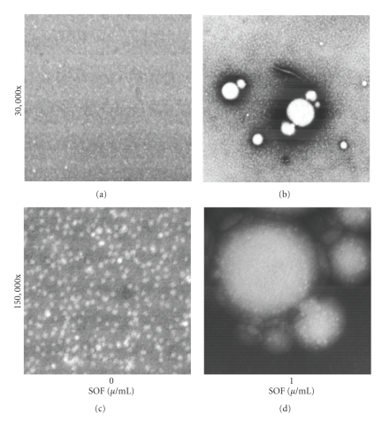
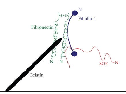
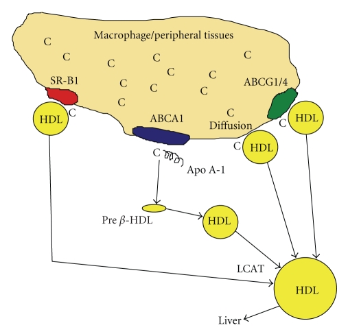
Serum opacity factor (SOF) is a virulence determinant expressed by a variety of streptococcal and staphylococcal species including both human and animal pathogens. SOF derives its name from its ability to opacify serum where it targets and disrupts the structure of high-density lipoproteins resulting in formation of large lipid vesicles that cause the serum to become cloudy. SOF is a multifunctional protein and in addition to its opacification activity, it binds to a number of host proteins that mediate adhesion of streptococci to host cells, and it plays a role in resistance to phagocytosis in human blood. This article will provide an overview of the structure and function of SOF, its role in the pathogenesis of streptococcal infections, its vaccine potential, its prevalence and distribution in bacteria, and the molecular mechanism whereby SOF opacifies serum and how an understanding of this mechanism may lead to therapies for reducing high-cholesterol concentrations in blood, a major risk factor for cardiovascular disease.


Figures

Figure 1
Figure 1
Opacification of human serum by SOF. Human serum was incubated overnight with either buffer control (left) or with 1 μg/ml of recombinant SOF (right).
Figure 2
Figure 2
A schematic indicating the location of functional domains within SOF. The yellow segments in the top bar indicate regions of SOF that are highly variable (vary in ≥60% of serotypes), black segments indicate highly conserved regions of SOF (conserved in ≥60% of serotypes. Only SOFs from S. pyogenes were used to generate this illustration. There are variations in the size of SOF from different serotypes but most are composed of ∼1050 amino acids including the leader sequence. The black lines signify the general location and size of the indicated domains. Abbreviations: vWFA: von Willebrand Factor A domain, MIDAS: metal ion-dependent adhesion site, Fn/Fgn: fibronectin/fibrinogen.
Figure 3
Figure 3
A comparison of the similarity between SOF from various serotypes of S. pyogenes and other streptococci (top). The bottom part of the figure illustrates the phylogenetic tree of SOF from different streptococci.
Figure 4
Figure 4
The predicted secondary structure of SOF is highly conserved among SOF from different serotypes. Only three serotypes were selected for comparison in order to have a manageable figure. The top figure indicates the degree of hydrophobicity/hydrophilicity and the bottom figure indicates location of helixes, turns, and B-sheets. The numbering of the amino acids is indicated.
Figure 5
Figure 5
Model of the opacification reaction of SOF with high-density lipoproteins (HDLs). SOF initiates the opacification reaction by binding to HDL. SOF is a heterodivalent fusogen that crosslinks two or more HDL particles and simultaneously induces the release of free apo A-I and promotes the fusion of the resultant particles culminating in the formation of a cholesterol-ester rich microemulsion (CERM) and neo-HDL. Neo-HDL is deficient in free cholesterol/cholesterol esters and rich in phospholipids and apo A-II. Some HDL particles also contain apo E, which is preferentially retained in CERM. Structures are not drawn to scale. CERM particles range from 100 to 500 nm in size whereas HDL particles are ∼8.5 nm [38]. Current data indicates one CERM particle contains cholesterol-esters from ∼400,000 HDL particles. It is these CERM particles that cause serum to become opaque due to their large size and insolubility in aqueous solutions.
Figure 6
Figure 6
Electron micrographs of HDL treated with SOF. Indicated concentrations of rSOF were incubated with human HDL overnight at 37°C, stained with 2% phosphotungstic acid, and electron micrographs taken at the indicated magnification.
Figure 7
Figure 7
Model of fibronectin. Fibronectin (Fn) is a large, dimeric glycoprotein with multiple functional domains. The 28 kDa N-terminal domain is the primary domain that interacts with most streptococci and staphylococci, but the collagen-binding domain may also interact with streptococci expressing protein F (or Sfb) [42]. SOF also binds to the N-terminal 28 kDa domain of Fn via the C-terminal, repeating peptide of SOF [40]. Fibulin-1 binds to the C-terminal, heparin-binding domain of Fn [43]. The interaction of streptococci with the N-terminal domain of soluble Fn is thought to induce a conformational change that exposes the RGD domain [44]. The RGD domain of Fn can then bind to integrins on the surface of host cells and tether the bacteria to the surface. Such interactions induce actin polymerization and promote internalization of the bacteria [45]. Streptococci and SOF can also bind to Fn that has already bound to surfaces. Hep: heparin, Fib: fibrin/fibrinogen.
Figure 8
Figure 8
Variants of the Mga regulon and their relationship to preferential colonization of host sites. Mga (multigene activator) is a positive regulator of a number of streptococcal genes. The most prominent of these are the family of M proteins whose genes are tandemly linked. sof and sfbx are bicistronic and are also regulated by Mga, but are located some distance away. emm encodes for M protein, mrp encodes M-related proteins, enn encodes an M-like protein that binds IgA, and scpa encodes a C5a peptidase. Some serotypes contain only mga, emm, and scpa (pattern A). Other serotypes contain one or more of the remaining genes (patterns B–E). S. pyogenes with Mga patterns A, B, and C primarily infect oral tissues, whereas strains with pattern D are predominantly found in tissues of the skin. S. pyogenes with a pattern E Mga regulon are found at both oral and skin sites. Figure derived from data and classification scheme of Bessen and co-workers [47, 48].
Figure 9
Figure 9
Model of quaternary complex between gelatin, fibronectin, fibulin-1, and SOF. Although SOF can bind to fibulin-1 independently of fibronectin, mixed binding experiments indicated that SOF binds much better to a complex of fibulin-1, fibronectin, and gelatin. Such complexes are possible because fibronectin contains independent binding sites for gelatin (collagen), fibulin-1, and SOF. Also SOF can react with fibulin-1 and fibronectin via independent binding domains. It is postulated that interactions between gelatin and fibronectin induces a conformational change in fibronectin that facilitates interactions with other ligands and SOF and enhances adhesion of streptococci to host surfaces. Reproduced from [49] with permission.
Figure 10
Figure 10
Reverse cholesterol transfer pathway (RCT). RCT is the major pathway for transfer of excess cholesterol (C) from peripheral tissues to the liver for disposal as bile. High levels of cholesterol can be toxic and the accumulation of cholesterol (C) in macrophages lining the blood vessels transforms these cells to foam cells leading to the development of plaque and atherosclerosis. Cholesterol is removed from these tissues and transferred to HDL by interactions with ATP binding cassette receptors ABCA1, ABCG1/4, and SR-B1 or by diffusion. The interaction between ABCA1 and apo A-I is the dominant pathway for removal of excess cholesterol from macrophages followed by an interaction between HDL and ABCG1. Together these two receptors account for about 70% of the efflux of excess cholesterol [86]. The size of HDL is modulated as its load of cholesterol increases and by interactions with various plasma factors such as lecithin cholesterol acyltransferase (LCAT) [87]. Free cholesterol removed from these tissues is esterified by LCAT and subsequently removed by liver cells by interactions with HDL. SOF enhances this process in several ways [88]. First, it releases free apo A-I, which is a better acceptor of free cholesterol than HDL. Secondly, it forms neo-HDL, a particle that is similar to pre-β HDL, which is also a better acceptor of cholesterol than HDL. Thirdly, SOF enhances cholesterol esterification, which may allow a more efficient uptake of cholesterol by the liver.

Similar articles

Cited by

References

    1. Ward HK, Rudd GV. Studies on haemolytic streptococci from human sources. Australian Journal of Experimental Biology & Medical Science. 1938;16:181–192.
    1. Krumwiede E. Studies on a lipoproteinase of group A streptococci. The Journal of Experimental Medicine. 1954;100(6):629–639. - PMC - PubMed
    1. Rowen R. The release of cholesterol esters from serum lipoproteins by extracts of certain group A streptococci. Journal of Experimental Medicine. 1961;114:807–823. - PMC - PubMed
    1. Rowen R, Martin J. Enhancement of cholesterol esterification in serum by an extract of group-A streptococcus. Biochimica et Biophysica Acta. 1963;70:396–405. - PubMed
    1. Saravani GA, Martin DR. Opacity factor from group A streptococci is an apoproteinase. FEMS Microbiology Letters. 1990;68(1-2):35–39. - PubMed

Publication types

LinkOut - more resources