

Cognitive Sparing during the Administration of Whole Brain Radiotherapy and Prophylactic Cranial Irradiation: Current Concepts and Approaches
- PMID: 20671962
- PMCID: PMC2910483
- DOI: 10.1155/2010/198208
Cognitive Sparing during the Administration of Whole Brain Radiotherapy and Prophylactic Cranial Irradiation: Current Concepts and Approaches
Abstract
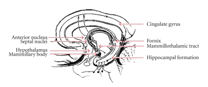
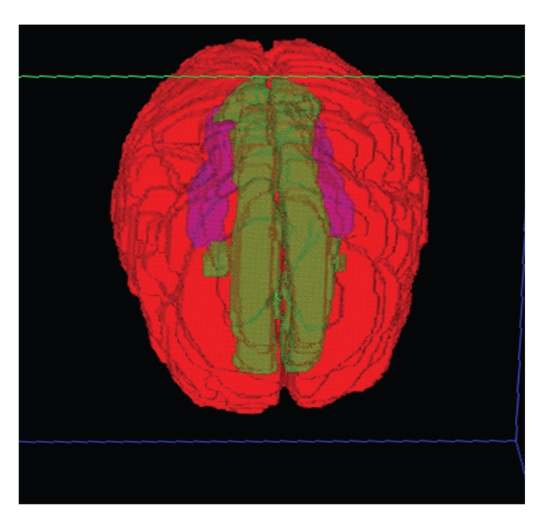
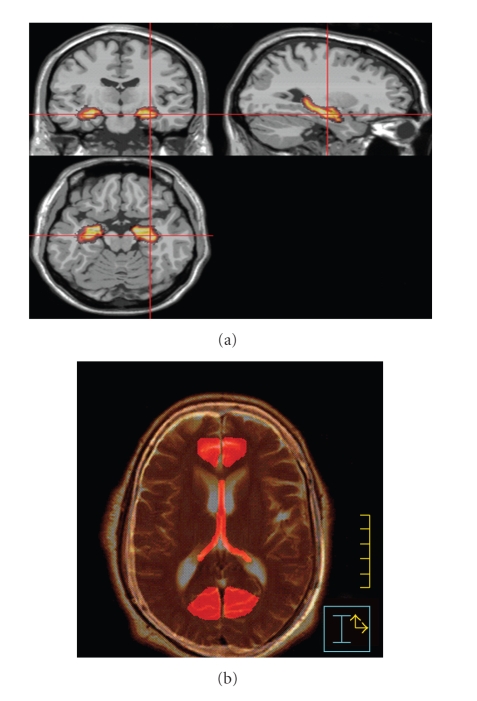
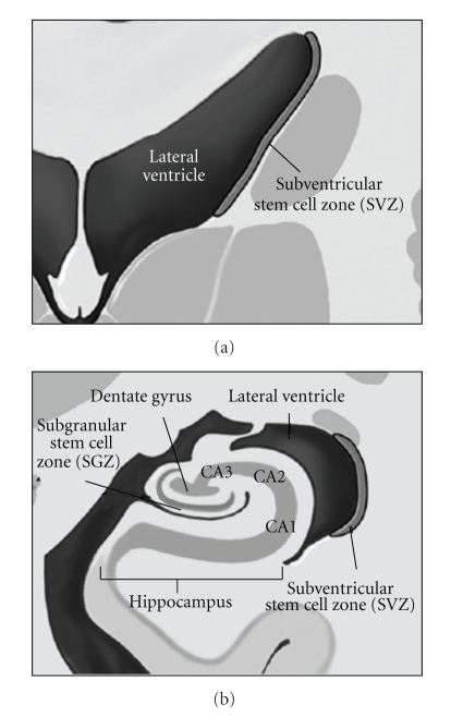
Whole brain radiotherapy (WBRT) for the palliation of metastases, or as prophylaxis to prevent intracranial metastases, can be associated with subacute and late decline in memory and other cognitive functions. Moreover, these changes are often increased in both frequency and severity when cranial irradiation is combined with the use of systemic or intrathecal chemotherapy. Approaches to preventing or reducing this toxicity include the use of stereotactic radiosurgery (SRS) instead of WBRT; dose reduction for PCI; exclusion of the limbic circuit, hippocampal formation, and/or neural stem cell regions of the brain during radiotherapy; avoidance of intrathecal and/or systemic chemotherapy during radiotherapy; the use of high-dose, systemic chemotherapy in lieu of WBRT. This review discusses these concepts in detail as well as providing both neuroanatomic and radiobiologic background relevant to these issues.
Figures
Similar articles
-
Cost-effectiveness of stereotactic radiosurgery with and without whole-brain radiotherapy for the treatment of newly diagnosed brain metastases.J Neurosurg. 2014 Dec;121 Suppl:84-90. doi: 10.3171/2014.7.GKS14972. J Neurosurg. 2014. PMID: 25434941
-
Randomised prospective phase II trial in multiple brain metastases comparing outcomes between hippocampal avoidance whole brain radiotherapy with or without simultaneous integrated boost: HA-SIB-WBRT study protocol.BMC Cancer. 2020 Oct 30;20(1):1045. doi: 10.1186/s12885-020-07565-y. BMC Cancer. 2020. PMID: 33126867 Free PMC article.
-
Outcome in patients with small cell lung cancer re-irradiated for brain metastases after prior prophylactic cranial irradiation.Lung Cancer. 2016 Nov;101:76-81. doi: 10.1016/j.lungcan.2016.09.010. Epub 2016 Sep 14. Lung Cancer. 2016. PMID: 27794411
-
Congress of Neurological Surgeons Systematic Review and Evidence-Based Guidelines on the Role of Whole Brain Radiation Therapy in Adults With Newly Diagnosed Metastatic Brain Tumors.Neurosurgery. 2019 Mar 1;84(3):E159-E162. doi: 10.1093/neuros/nyy541. Neurosurgery. 2019. PMID: 30629211
-
Strategies to Preserve Cognition in Patients With Brain Metastases: A Review.Front Oncol. 2018 Oct 9;8:415. doi: 10.3389/fonc.2018.00415. eCollection 2018. Front Oncol. 2018. PMID: 30356657 Free PMC article. Review.
Cited by
-
Glitches in the brain: the dangerous relationship between radiotherapy and brain fog.Front Cell Neurosci. 2024 Mar 7;18:1328361. doi: 10.3389/fncel.2024.1328361. eCollection 2024. Front Cell Neurosci. 2024. PMID: 38515789 Free PMC article. Review.
-
Cell cycle inhibition without disruption of neurogenesis is a strategy for treatment of aberrant cell cycle diseases: an update.ScientificWorldJournal. 2012;2012:491737. doi: 10.1100/2012/491737. Epub 2012 Apr 1. ScientificWorldJournal. 2012. PMID: 22547985 Free PMC article. Review.
-
Future directions in treatment of brain metastases.Surg Neurol Int. 2013 May 2;4(Suppl 4):S220-30. doi: 10.4103/2152-7806.111299. Print 2013. Surg Neurol Int. 2013. PMID: 23717793 Free PMC article.
-
Non-coplanar volumetric-modulated arc therapy (VMAT) for craniopharyngiomas reduces radiation doses to the bilateral hippocampus: a planning study comparing dynamic conformal arc therapy, coplanar VMAT, and non-coplanar VMAT.Radiat Oncol. 2016 Jun 23;11:86. doi: 10.1186/s13014-016-0659-x. Radiat Oncol. 2016. PMID: 27338798 Free PMC article.
-
Innovative therapeutic strategies in the treatment of brain metastases.Int J Mol Sci. 2013 Jan 22;14(1):2135-74. doi: 10.3390/ijms14012135. Int J Mol Sci. 2013. PMID: 23340652 Free PMC article.
References
-
- Ewend MG, Elbabaa S, Carey LA. Current treatment paradigms for the management of patients with brain metastases. Neurosurgery. 2005;57(5):S66–S77. - PubMed
-
- Mehta MP, Khuntia D. Current strategies in whole-brain radiation therapy for brain metastases. Neurosurgery. 2005;57(5):S33–S44. - PubMed
-
- Peacock KH, Lesser GJ. Current therapeutic approaches in patients with brain metastases. Current Treatment Options in Oncology. 2006;7(6):479–489. - PubMed
-
- Tsao MN, Lloyd N, Wong R, Chow E, Rakovitch E, Laperriere N. Whole brain radiotherapy for the treatment of multiple brain metastases. Cochrane Database of Systematic Reviews. 2006;3 Article ID CD003869. - PubMed
-
- Gaspar L, Scott C, Rotman M, et al. Recursive Partitioning Analysis (RPA) of prognostic factors in three Radiation Therapy Oncology Group (RTOG) brain metastases trials. International Journal of Radiation Oncology Biology Physics. 1997;37(4):745–751. - PubMed
LinkOut - more resources
Full Text Sources
Medical
Miscellaneous
