. 2010:2010:139167.
doi: 10.1155/2010/139167. Epub 2010 Jun 30.

Desmosomal molecules in and out of adhering junctions: normal and diseased States of epidermal, cardiac and mesenchymally derived cells

Affiliations

Desmosomal molecules in and out of adhering junctions: normal and diseased States of epidermal, cardiac and mesenchymally derived cells

Sebastian Pieperhoff et al. Dermatol Res Pract. 2010.

Abstract

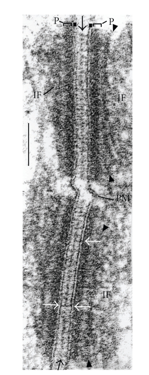
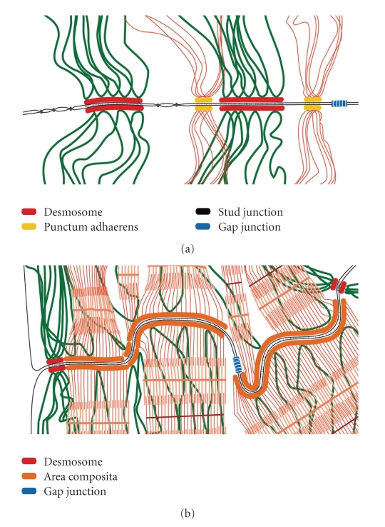
Current cell biology textbooks mention only two kinds of cell-to-cell adhering junctions coated with the cytoplasmic plaques: the desmosomes (maculae adhaerentes), anchoring intermediate-sized filaments (IFs), and the actin microfilament-anchoring adherens junctions (AJs), including both punctate (puncta adhaerentia) and elongate (fasciae adhaerentes) structures. In addition, however, a series of other junction types has been identified and characterized which contain desmosomal molecules but do not fit the definition of desmosomes. Of these special cell-cell junctions containing desmosomal glycoproteins or proteins we review the composite junctions (areae compositae) connecting the cardiomyocytes of mature mammalian hearts and their importance in relation to human arrhythmogenic cardiomyopathies. We also emphasize the various plakophilin-2-positive plaques in AJs (coniunctiones adhaerentes) connecting proliferatively active mesenchymally-derived cells, including interstitial cells of the heart and several soft tissue tumor cell types. Moreover, desmoplakin has also been recognized as a constituent of the plaques of the complexus adhaerentes connecting certain lymphatic endothelial cells. Finally, we emphasize the occurrence of the desmosomal transmembrane glycoprotein, desmoglein Dsg2, out of the context of any junction as dispersed cell surface molecules in certain types of melanoma cells and melanocytes. This broadening of our knowledge on the diversity of AJ structures indicates that it may still be too premature to close the textbook chapters on cell-cell junctions.


Figures

Figure 1
Figure 1
High-magnification electron micrograph of two cross sectioned desmosomal structures connecting stratum spinosum cells of human fetal (20 wk) foot-sole epidermis. Brackets and label “P”: cytoplasmic dense plaque; black arrows: midline structure; white arrows: trilaminar “unit membrane” structure of the plasma membrane; arrowheads (top and bottom): secondary dense layer of the plaque; IF: intermediate-sized filaments (for further details see [7]); PM: plasma membrane. Bar: 0.1 μm.
Figure 2
Figure 2
Immunofluorescence microscopy of cryostat sections through fish and amphibian skin, showing the localization of desmosomal components in all layers of the epidermis. (a) Immunofluorescence labeling of plakoglobin (red), in addition to nuclear DAPI staining (blue), in a section through the paraffin-embedded skin of an eel (Anguilla anguilla). The section is shown after antigen retrieval and immunoreaction (for methods see [8]) using a monoclonal mouse plakoglobin antibody (mAb PG 5.1; Progen Biotechnik, Heidelberg, Germany). Note the distinct basal layer (stratum basale) of the epidermis and the relatively large club shaped cells (“club cells”) as well as the mucous goblet cells and the dense-packed apical cell layers. (b) Immunofluorescence labeling of desmoplakin on a cryostat section through the skin of the frog, Rana pipiens, using mAb DP447 (Progen Biotechnik). Note the continuous pattern of very closely spaced, finely punctate staining of the epidermal desmosomes. Bl: basal layer; cc: club cells; gc: goblet cells; sec: superficial epithelial cells (for histological terminology see [8]). Bar in (a): 100 μm; bar in (b): 50 μm.
Figure 3
Figure 3
Micrographs showing the immunolocalization of the desmosomal plaque protein, desmoplakin, on cryostat sections through the murine myocardium. (a) Immunofluorescence micrograph showing the localization of desmoplakin (red) in all composite junction structures of the intercalated disks (IDs). (b) Immunofluorescence micrograph showing desmoplakin (red) labeling of the composite junctions on the background of phase-contrast optics. (c) Merged image showing plakophilin-2 (red), β-catenin (green) and nuclear DAPI-staining (blue), on an interference contrast microscopy background. Composite junctions (CJs) are characterized by their yellow merge colour whereas the zonulae adhaerentes and other AJs of the capillary endothelial cell layers here show only β-catenin-positivity. Bar in (a): 100 μm; bar in (b): 50 μm; bar in (c): 25 μm.
Figure 4
Figure 4
Schematic comparison of the different principles of organization of cell-cell junctions containing desmosomal proteins in mammalian epidermal and myocardiac tissue. (a) Junction organizations between epidermal keratinocytes as observed in the stratum spinosum. Note the densely spaced desmosomes (red plaques), which anchor bundles of intermediate sized filaments containing keratins (dark-green). Smaller adherens junctions (puncta adhaerentia, yellow plaques) anchor actin-microfilament bundles (red filaments). Furthermore, special types of tight junction-like structures, the small “stud junctions” (black dots), and channel-like connexin paracrystals (gap junctions, blue) are also generally found. For details see [29, 30]. (b) The area composita (composite junction, orange) structures of intercalated disks (ID) connecting cardiomyocytes of an adult mammalian heart. This amalgamated type of adhering junction is characterized by a mixture of typical desmosomal and AJ molecules. This kind of composite junction is the predominating adhering junction structure in the ID of the adult mammalian heart.
Figure 5
Figure 5
Immunoelectron microscopy of ultrathin sections through bovine myocardium (a) and Purkinje fibers ((b)–(d)) using antibodies against desmoplakin. (a) Immunogold labeling of a typical extended area composita structure in an intercalated disk (ID) of adult cardiomyocytes. (b) By contrast, note the relatively small desmoplakin-positive junctions that connect cells of the Purkinje fiber conductive system ((d), desmosomes; arrowheads denote some particularly small desmosome-like junction structures with asymmetric labeling). The desmoplakin-rich plaques of such junctions are very intensely labeled. (c) The morphology and the relatively close packing of the major type of junctions are similar to those of the area composita structures of adult mammalian cardiomyocytes. (d) High-magnification immunoelectron micrograph of a composite junction. Note the very close near parallel association of intermediate-sized filaments (some are denoted by arrows) with junctional plaques. Bars in (a) and (b): 1 μm; bar in (c): 0.25 μm.
Figure 6
Figure 6
Double-label immunofluorescence micrographs, showing the localization of the “desmosomal protein”, plakophilin-2, in mesenchymally derived cultures of valvular interstitial cells (VICs). (a), (a′) Phase contrast and immunofluorescence micrograph showing ovine VICs forming clusters of AJs positive for the typical desmosomal plaque component plakophilin-2 (red; vimentin filaments are labeled in green). (b)–(d) Represent merged images of ovine VICs, showing colocalization staining (yellow) of plakophilin-2 (red) with typical AJ proteins such as cadherin-11 (green, (b)), β-catenin (green, (c)), protein p120 (green, (d)) all on a phase contrast background. (e) Human VICs exhibit similar colocalization of plakophilin-2 (red) here with N-cadherin (green) as shown by the yellow merge colour (for further details see [23]). Bar in (a): 25 μm; bar in (b): 30 μm; bars in (c) and (d): 20 μm; bar in E: 10 μm.

Similar articles

Cited by

References

    1. Farquhar MG, Palade GE. Junctional complexes in various epithelia. The Journal of Cell Biology. 1963;17:375–412. - PMC - PubMed
    1. Drochmans P, Freudenstein C, Wanson JC, et al. Structure and biochemical composition of desmosomes and tonofilaments isolated from calf muzzle epidermis. Journal of Cell Biology. 1978;79:427–443. - PMC - PubMed
    1. Cowin P, Franke WW, Grund C, Kapprell HP, Kartenbeck J. The desmosome-intermediate filament complex. In: Edelman GM, Thiery JP, editors. The Cell in Contact. New York, NY, USA: John Wiley & Sons; 1985. pp. 427–460.
    1. Godsel LM, Getsios S, Huen AC, Green KJ. The molecular composition and function of desmosomes. In: Behrens J, Nelson WJ, editors. Cell Adhesion. Heidelberg, Germany: Springer; 2004. pp. 137–193. - PubMed
    1. Garrod D, Chidgey M. Desmosome structure, composition and function. Biochimica et Biophysica Acta. 2008;1778(3):572–587. - PubMed

LinkOut - more resources