

Indeterminate cell histiocytosis in association with acute myeloid leukemia
- PMID: 20672000
- PMCID: PMC2905718
- DOI: 10.1155/2010/569345
Indeterminate cell histiocytosis in association with acute myeloid leukemia
Abstract
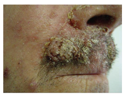
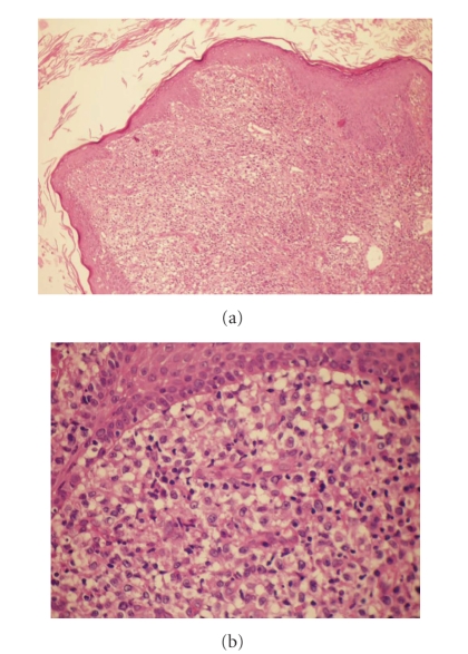
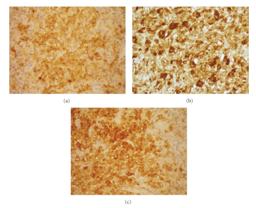
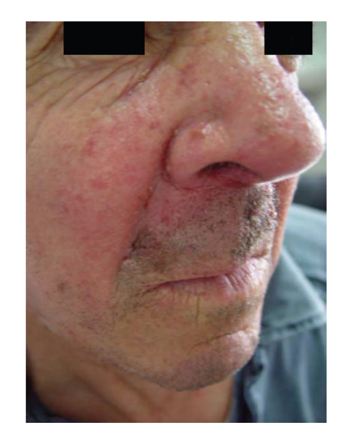
Indeterminate cell histiocytosis (ICH) is a rare proliferative disorder, in which the predominant cells share morphologic and immunophenotypic features from both Langerhans and non-Langerhans cell histiocytosis. We describe a 62-year-old man presenting a 2-month history of firm nodular lesions on the upper lip. Histopathology, immunohistochemical, and ultrastructural analysis showed typical findings of ICH. The patient was treated with thalidomide and almost complete regression of the lesions was reached within 7 months. Nevertheless, one month after remission, he developed an acute myeloid leukemia of the subtype monocytic leukemia (M5). The patient's condition rapidly worsened and he died due to a respiratory failure four weeks later. We present this case because apart of being rare it joins the effectiveness of thalidomide and the association with an acute monocytic leukemia. A review of the literature is made.
Figures
References
-
- Vener C, Soligo D, Berti E, et al. Indeterminate cell histiocytosis in association with later occurrence of acute myeloblastic leukaemia. British Journal of Dermatology. 2007;156(6):1357–1361. - PubMed
-
- Ratzinger G, Burgdorf WHC, Metze D, Zeiger BG, Zelger B. Indeterminate cell histiocytosis: fact or fiction? Journal of Cutaneous Pathology. 2005;32(8):552–560. - PubMed
-
- Newman B, Hu W, Nigro K, Gilliam AC. Aggressive histiocytic disorders that can involvetheskin. Journal of the American Academy of Dermatology. 2007;56(2):302–316. - PubMed
-
- Dzierzak E. Embryonic beginnings of definitive hematopoietic stem cells. Annals of the New York Academy of Sciences. 1999;872:256–264. - PubMed
-
- Sidoroff A, Zelger B, Steiner H, Smith N. Indeterminate cell histiocytosis—a clinicopathological entity with features of both X- and non-X histiocytosis. British Journal of Dermatology. 1996;134(3):525–532. - PubMed
Publication types
LinkOut - more resources
Full Text Sources
