

Chondrogenesis and mineralization during in vitro culture of human mesenchymal stem cells on three-dimensional woven scaffolds
- PMID: 20673022
- PMCID: PMC2991213
- DOI: 10.1089/ten.TEA.2010.0190
Chondrogenesis and mineralization during in vitro culture of human mesenchymal stem cells on three-dimensional woven scaffolds
Abstract
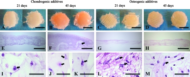
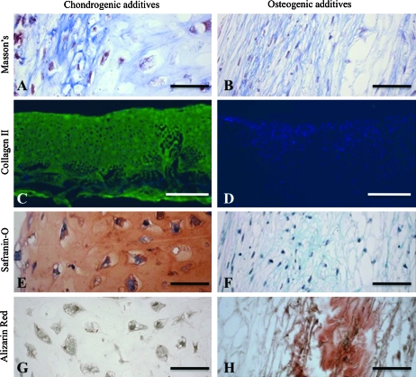
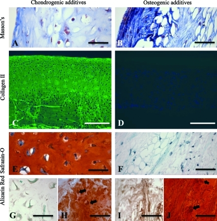
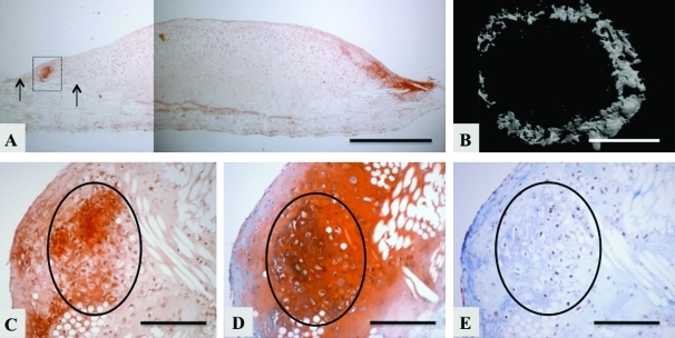
Human mesenchymal stem cells (hMSCs) and three-dimensional (3D) woven poly(ɛ-caprolactone) (PCL) scaffolds are promising tools for skeletal tissue engineering. We hypothesized that in vitro culture duration and medium additives can individually and interactively influence the structure, composition, mechanical, and molecular properties of engineered tissues based on hMSCs and 3D poly(ɛ-caprolactone). Bone marrow hMSCs were suspended in collagen gel, seeded on scaffolds, and cultured for 1, 21, or 45 days under chondrogenic and/or osteogenic conditions. Structure, composition, biomechanics, and gene expression were analyzed. In chondrogenic medium, cartilaginous tissue formed by day 21, and hypertrophic mineralization was observed in the newly formed extracellular matrix at the interface with underlying scaffold by day 45. Glycosaminoglycan, hydroxyproline, and calcium contents, and alkaline phosphatase activity depended on culture duration and medium additives, with significant interactive effects (all p < 0.0001). The 45-day constructs exhibited mechanical properties on the order of magnitude of native articular cartilage (aggregate, Young's, and shear moduli of 0.15, 0.12, and 0.033 MPa, respectively). Gene expression was characteristic of chondrogenesis and endochondral bone formation, with sequential regulation of Sox-9, collagen type II, aggrecan, core binding factor alpha 1 (Cbfα1)/Runx2, bone sialoprotein, bone morphogenetic protein-2, and osteocalcin. In contrast, osteogenic medium produced limited osteogenesis. Long-term culture of hMSC on 3D scaffolds resulted in chondrogenesis and regional mineralization at the interface between soft, newly formed engineered cartilage, and stiffer underlying scaffold. These findings merit consideration when developing grafts for osteochondral defect repair.
Figures
Similar articles
-
The inhibition by interleukin 1 of MSC chondrogenesis and the development of biomechanical properties in biomimetic 3D woven PCL scaffolds.Biomaterials. 2012 Dec;33(35):8967-74. doi: 10.1016/j.biomaterials.2012.08.045. Epub 2012 Sep 19. Biomaterials. 2012. PMID: 22999467 Free PMC article.
-
In vitro generation of mechanically functional cartilage grafts based on adult human stem cells and 3D-woven poly(epsilon-caprolactone) scaffolds.Biomaterials. 2010 Mar;31(8):2193-200. doi: 10.1016/j.biomaterials.2009.11.092. Epub 2010 Jan 19. Biomaterials. 2010. PMID: 20034665 Free PMC article.
-
Spatial regulation of human mesenchymal stem cell differentiation in engineered osteochondral constructs: effects of pre-differentiation, soluble factors and medium perfusion.Osteoarthritis Cartilage. 2010 May;18(5):714-23. doi: 10.1016/j.joca.2010.01.008. Epub 2010 Feb 6. Osteoarthritis Cartilage. 2010. PMID: 20175974 Free PMC article.
-
Genetic Engineering of Mesenchymal Stem Cells for Differential Matrix Deposition on 3D Woven Scaffolds.Tissue Eng Part A. 2018 Oct;24(19-20):1531-1544. doi: 10.1089/ten.TEA.2017.0510. Epub 2018 Jul 13. Tissue Eng Part A. 2018. PMID: 29756533 Free PMC article.
-
Investigation of multiphasic 3D-bioplotted scaffolds for site-specific chondrogenic and osteogenic differentiation of human adipose-derived stem cells for osteochondral tissue engineering applications.J Biomed Mater Res B Appl Biomater. 2020 Jul;108(5):2017-2030. doi: 10.1002/jbm.b.34542. Epub 2019 Dec 27. J Biomed Mater Res B Appl Biomater. 2020. PMID: 31880408 Free PMC article.
Cited by
-
Textile Technologies and Tissue Engineering: A Path Toward Organ Weaving.Adv Healthc Mater. 2016 Apr 6;5(7):751-66. doi: 10.1002/adhm.201500517. Epub 2016 Feb 29. Adv Healthc Mater. 2016. PMID: 26924450 Free PMC article. Review.
-
TGF-β3-induced chondrogenesis in co-cultures of chondrocytes and mesenchymal stem cells on biodegradable scaffolds.Biomaterials. 2014 Jan;35(1):123-32. doi: 10.1016/j.biomaterials.2013.09.086. Epub 2013 Oct 11. Biomaterials. 2014. PMID: 24125773 Free PMC article.
-
The inhibition by interleukin 1 of MSC chondrogenesis and the development of biomechanical properties in biomimetic 3D woven PCL scaffolds.Biomaterials. 2012 Dec;33(35):8967-74. doi: 10.1016/j.biomaterials.2012.08.045. Epub 2012 Sep 19. Biomaterials. 2012. PMID: 22999467 Free PMC article.
-
The use of mesenchymal stem cells for cartilage repair and regeneration: a systematic review.J Orthop Surg Res. 2017 Mar 9;12(1):39. doi: 10.1186/s13018-017-0534-y. J Orthop Surg Res. 2017. PMID: 28279182 Free PMC article.
-
Multimodal evaluation of tissue-engineered cartilage.J Med Biol Eng. 2013 Feb 1;33(1):1-16. doi: 10.5405/jmbe.1254. J Med Biol Eng. 2013. PMID: 23606823 Free PMC article.
References
-
- Gao J. Dennis J.E. Solchaga L.A. Awadallah A.S. Goldberg V.M. Caplan A.I. Tissue-engineered fabrication of an osteochondral composite graft using rat bone marrow-derived mesenchymal stem cells. Tissue Eng. 2001;7:363. - PubMed
-
- Schaefer D. Martin I. Jundt G. Seidel J. Heberer M. Grodzinsky A.J. Bergin I. Vunjak-Novakovic G. Freed L.E. Tissue engineered composites for the repair of large osteochondral defects. Arthritis Rheum. 2002;46:2524. - PubMed
-
- Sherwood J.K. Riley S.L. Palazzolo R. Brown S.C. Monkhouse D.C. Coates M. Griffith L.G. Landeen L.K. Ratcliffe A. A three-dimensional osteochondral composite scaffold for articular cartilage repair. Biomaterials. 2002;23:4739. - PubMed
Publication types
MeSH terms
Grants and funding
LinkOut - more resources
Full Text Sources
Other Literature Sources
Research Materials
Miscellaneous
