Review
. 2011 May 15;14(10):1889-903.
doi: 10.1089/ars.2010.3474. Epub 2010 Dec 4.

Significance of brain tissue oxygenation and the arachidonic acid cascade in stroke

Affiliations
Review

Significance of brain tissue oxygenation and the arachidonic acid cascade in stroke

Cameron Rink et al. Antioxid Redox Signal. .

Abstract

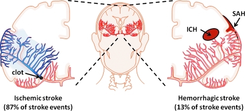
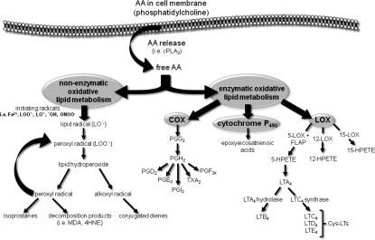
The significance of the hypoxia component of stroke injury is highlighted by hypermetabolic brain tissue enriched with arachidonic acid (AA), a 22:6n-3 polyunsaturated fatty acid. In an ischemic stroke environment in which cerebral blood flow is arrested, oxygen-starved brain tissue initiates the rapid cleavage of AA from the membrane phospholipid bilayer. Once free, AA undergoes both enzyme-independent and enzyme-mediated oxidative metabolism, resulting in the formation of number of biologically active metabolites which themselves contribute to pathological stroke outcomes. This review is intended to examine two divergent roles of molecular dioxygen in brain tissue as (1) a substrate for life-sustaining homeostatic metabolism of glucose and (2) a substrate for pathogenic metabolism of AA under conditions of stroke. Recent developments in research concerning supplemental oxygen therapy as an intervention to correct the hypoxic component of stroke injury are discussed.


Figures

FIG. 1.
FIG. 1.
Stroke etiology. Stroke is a general term used to define pathological conditions in which cerebral blood flow is disrupted. The cause of stroke can be classified as one of two etiological origins: ischemic (left) and hemorrhagic (right). Ischemic stroke describes the arrest of cerebral blood flow by narrowing and blockage of a cerebral artery (thrombosis) or by a peripheral artery clot (emboli) traveling to and blocking a cerebral artery. Hemorrhagic stroke describes the rupture of a cerebral artery in intracerebral space (ICH) or subarachnoid space (SAH). (To see this illustration in color the reader is referred to the web version of this article at www.liebertonline.com/ars).
FIG. 2.
FIG. 2.
Characterization of the ischemic penumbra in focal ischemic stroke. In a focal ischemic stroke event, brain tissue oxygenation increases toward baseline (normoxia, green) with distance from the ischemic core (red). Ischemic penumbra and core are delineated by quantitative molecular and physiological differences. (To see this illustration in color the reader is referred to the web version of this article at www.liebertonline.com/ars).
FIG. 3.
FIG. 3.
Stroke-induced hypoxia mediates oxidative metabolism of arachidonic acid. Following release from the lipid membrane bi-layer by oxygen-sensitive phospholipase A2 (PLA2), arachidonic acid (AA) undergoes oxidative metabolism under enzymatic and nonenzymatic mechanisms. FLAP, 5-LOX activating protein; 4HNE, 4-hydroxynonenal; 5-HPETE, 5-hydroperoxyeicosatetraenoic acid; 12-HPETE, 12-hydroperoxyeicosatetraenoic acid; 15-HPETE, 15-hydroperoxyeicosatetraenoic acid; 5-LOX, 5-lipoxygenase; 12-LOX, 12-lipoxygenase; 15-LOX, 15-lipoxygenase; LTA4, leukotriene A4; LTB4, leukotriene B4; LTC4, leukotriene C4; LTD4, leukotriene D4; MDA, malondialdehyde; PGD2, prostaglandin D2; PGF2a, prostaglandin F2a; PGG2, prostaglandin G2; PGH2, prostaglandin H2; PGI2, prostaglandin I2; TXA2, thromboxane A2.
FIG. 4.
FIG. 4.
Hyperbaric oxygen therapy reduces during, exacerbates at reperfusion, acute ischemic stroke infarct volume. (A) Representative T2-weighted MR images were acquired 48 h after MCAO in rodents kept in room air during ischemia (RA, control), or receiving hyperbaric oxygen (100% O2 at 2 ATA) during ischemia (iHBO) or immediately following reperfusion (rHBO). Color look up table applied—shift from blue to red denotes edema and stroke-induced infarct. Three-dimensional reconstruction of coronal slices permits visualization of the brain from dorsal and oblique positions. HBO during MCAO significantly decreased stroke-induced lesion volume in iHBO animals, while in rHBO animals is increased lesion volume. (B) Percent hemispherical lesion volume corrected for edema >5 *p < 0.05 vs RA, †p < 0.05 vs iHBO. (To see this illustration in color the reader is referred to the web version of this article at www.liebertonline.com/ars).
FIG. 5.
FIG. 5.
HBO corrects stroke-induced hypoxia during middle cerebral artery occlusion in rodents. (A) Oxygen-sensitive EPR oximetry probe was implanted in the ischemic penumbra of rodents in the week prior to MCAO using stereotaxic coordinates −0.1 mm bregma, +2.0 mm lateral, −1.0 mm dorsal. Placement of the probe was verified using MRI. (B) EPR spectra were acquired prior to MCAO to characterize normal cortical pO2 (baseline). After MCAO-induced focal ischemia, brain pO2 in the ischemic penumbra significantly dropped to ∼ 50% of baseline level. Correction of stroke-induced hypoxia was achieved with HBO during ischemia in the penumbra zone. Increased noise in EPR signal was encountered in the HBO group due to greater distance between probe and resonator as a result of the hyperbaric chamber. Representative EPR spectra shown. N ≥ 3, *p < 0.05 vs baseline, p < 0.05 vs ischemia.

References

    1. Abramovitz M. Wong E. Cox ME. Richardson CD. Li C. Vickers PJ. 5-lipoxygenase-activating protein stimulates the utilization of arachidonic acid by 5-lipoxygenase. Eur J Biochem. 1993;215:105–111. - PubMed
    1. Acker T. Acker H. Cellular oxygen sensing need in CNS function: Physiological and pathological implications. J Exp Biol. 2004;207:3171–3188. - PubMed
    1. Adibhatla RM. Hatcher JF. Dempsey RJ. Phospholipase A2, hydroxyl radicals, and lipid peroxidation in transient cerebral ischemia. Antioxid Redox Signal. 2003;5:647–654. - PubMed
    1. Adibhatla RM. Hatcher JF. Dempsey RJ. Lipids and lipidomics in brain injury and diseases. AAPS J. 2006;8:E314–21. - PMC - PubMed
    1. Adibhatla RM. Hatcher JF. Larsen EC. Chen X. Sun D. Tsao FH. CDP-choline significantly restores phosphatidylcholine levels by differentially affecting phospholipase A2 and CTP: Phosphocholine cytidylyltransferase after stroke. J Biol Chem. 2006;281:6718–6725. - PubMed

Publication types