. 2010 Dec;131(4):549-55.
doi: 10.1111/j.1365-2567.2010.03327.x.

Maternally transferred anti-factor VIII IgG reduce the anti-factor VIII humoral immune response in factor VIII-deficient mice

Affiliations

Maternally transferred anti-factor VIII IgG reduce the anti-factor VIII humoral immune response in factor VIII-deficient mice

Yann Meslier et al. Immunology. 2010 Dec.

Abstract

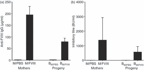
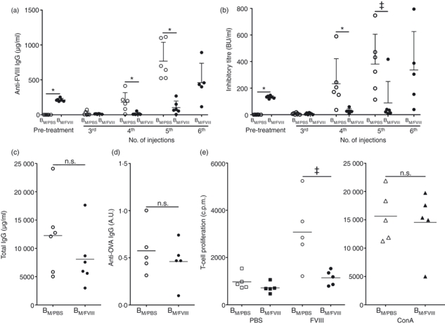
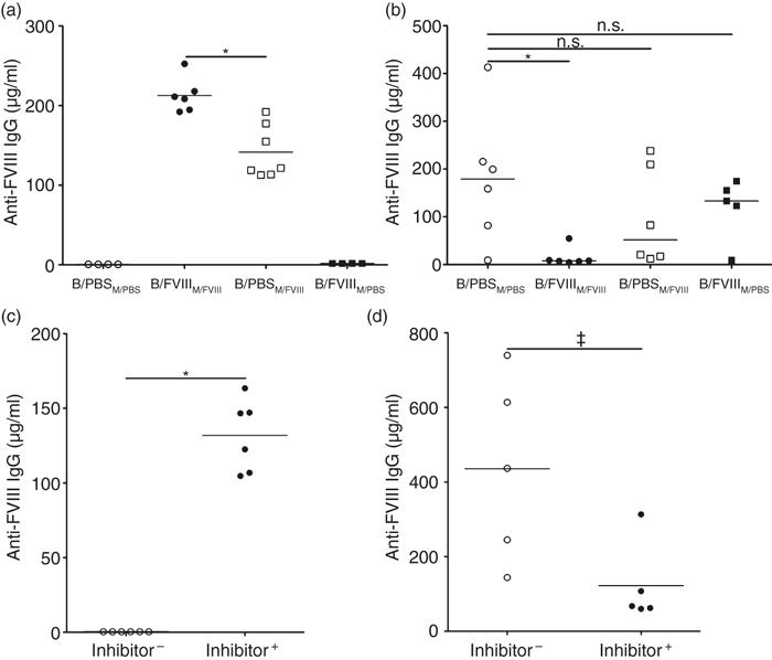
Replacement therapy with exogenous factor VIII (FVIII) to treat haemorrhages or used in prophylaxis induces inhibitory anti-FVIII immunoglobulin G (IgG) in some patients with haemophilia A. Therapeutic strategies to prevent the onset of the deleterious anti-FVIII immune response are still lacking. Maternal IgG is transferred to the offspring during fetal and neonatal life. While protecting the offspring from bacterial and viral infections, maternal IgG may alter the repertoires of T and B lymphocytes, and may impair vaccination in early infancy. Using haemophilic mice, we demonstrate that the transfer of maternal anti-FVIII IgG modulates the onset of anti-FVIII inhibitory IgG in early adulthood. The protective effect is reproduced upon reconstitution of naive mice with anti-FVIII IgG, suggesting that the reduced ability to mount an anti-FVIII immune response is the result of an interference between circulating anti-FVIII IgG and the administered FVIII rather than to a profound remodelling of lymphocyte repertoires occurring during the ontogeny of the immune system.


Figures

Figure 1
Figure 1
Transfer of maternal anti-factor VIII (FVIII) immunoglobulin G (IgG) to the fetus during pregnancy. Factor VIII-deficient female mice were administered intravenously four times once a week with 1 IU of human recombinant FVIII (M/FVIII) or with phosphate-buffered saline (M/PBS). The mice were bred before the last FVIII injection. At the time of delivery, blood was collected from newborns from FVIII-treated mothers (BM/FVIII) and from PBS-treated mothers (BM/PBS) and from their respective mothers. The titres of anti-FVIII IgG were measured in serum by enzyme-linked immunosorbent assay (a) and by Bethesda assay (b). Titres are as Bethesda Units/ml (three mice for each group).
Figure 2
Figure 2
Effect of maternally transferred anti-factor VIII (FVIII) immunoglobulin G (IgG) on the anti-FVIII immune response in the offspring. (a,b) Reduction of the humoral anti-FVIII immune response upon transfer of maternal anti-FVIII IgG. From 8 weeks of age onwards, the progeny from FVIII-treated mothers (BM/FVIII, closed circles) and from phosphate-buffered saline (PBS) -treated mothers (BM/PBS, open circles) were treated with FVIII once a week. Blood was collected from the offspring before FVIII treatment (8 weeks old, Pre-treatment) to assess the residual levels of anti-FVIII maternal IgG. The progeny were also bled after the third, fourth, fifth and sixth FVIII administrations. The graph depicts the levels of anti-FVIII IgG measured in serum by enzyme-linked immunosorbent assay (ELISA) (a) and the inhibitory activity in serum (b). (c) Levels of total IgG in the serum of offsprings from FVIII-treated mothers (BM/FVIII, closed circles) and PBS-treated mothers (BM/PBS, open circles), after the fourth FVIII administration, as measured by ELISA. (d) Anti-ovalbumin (OVA) immune response in the progeny from FVIII-treated mothers. The progeny of FVIII- (BM/FVIII, closed circles) and PBS-treated mothers (BM/PBS, open circles) was immunized at 8 weeks with OVA. Titres of anti-OVA IgG were measured in the serum of the mice by ELISA. (e) Reduction of the FVIII-specific T-cell proliferation upon transfer of maternal anti-FVIII IgG. Splenocytes were prepared from the progeny of FVIII-treated mothers (BM/FVIII, closed symbols) and PBS-treated mothers (BM/PBS, open symbols). Splenocytes were incubated alone (squares), with FVIII (circles) or concanavalin A (Con A; triangles). Cell proliferation was assessed by incorporation of [3H]thymidine, and is expressed as counts per minute (c.p.m.). Statistical significance in differences was assessed using the non-parametric Mann–Whitney U-test (*P < 0·01; ‡P < 0·05). Data are representative of two independent experiments.
Figure 3
Figure 3
Immunomodulatory effects of anti-factor VIII (FVIII) immunoglobulin G (IgG). (a,b). Comparative immunomodulatory effects of trans-placental and trans-epithelial transfer of anti-FVIII maternal IgG. At the time of delivery, the progeny from FVIII-treated mothers were transferred to phosphate-buffered saline (PBS) -treated mothers (referred to as B/FVIIIM/PBS), while the progeny from PBS-treated mothers were transferred to FVIII-treated mothers (referred to as B/PBSM/FVIII). In parallel, the progeny from FVIII- and PBS-treated mothers were kept with their respective mothers (referred to as B/FVIIIM/FVIII and B/PBSM/PBS, respectively). From 8 weeks of age onwards, the different groups of offspring were treated with 1 IU FVIII once a week for 4 weeks. Blood was collected before the first FVIII administration (a) to assess the titres of residual maternal anti-FVIII IgG, and 5 days after the fourth FVIII administration (b) to assess the titres of elicited anti-FVIII IgG. Statistical significance in differences was assessed using the non-parametric Mann–Whitney U-test (*P < 0·01). (c). Naive FVIII-deficient mice were reconstituted with IgG purified from the serum of FVIII-treated deficient mice (referred to as ‘Inhibitor+’ mice, full circles, 10 mg of total IgG containing 200 μg of anti-FVIII IgG ESH8-equivalent). As control, naive FVIII-deficient mice were given 10 mg of IgG purified from the serum of naive FVIII-deficient mice (referred to as ‘Inhibitor−’ mice, empty circles). The graph depicts the residual anti-FVIII IgG titres, as measured in serum by enzyme-linked immunosorbent assay (ELISA) 7 hr after infusion. (d) Reconstituted Inhibitor+ and Inhibitor− mice were given 1 IU of FVIII once a week for 6 weeks. The graph depicts the titres of anti-FVIII IgG, as measured by ELISA in the serum collected 5 days after the last FVIII administration. Statistical significance in differences was assessed using the non-parametric Mann–Whitney U-test (*P < 0·01; ‡P < 0·05).

Similar articles

Cited by

References

    1. Mariani G, Ghirardini A, Bellocco R. Immune tolerance in hemophilia: principal results from the international registry. Thromb Haemost. 1994;72:155–8. - PubMed
    1. Lacroix-Desmazes S, Navarrete AM, Andre S, Bayry J, Kaveri SV, Dasgupta S. Dynamics of factor VIII interactions determine its immunologic fate in hemophilia A. Blood. 2008;112:240–9. - PubMed
    1. Kurnik K, Bidlingmaier C, Engl W, Chehadeh H, Reipert B, Auerswald G. New early prophylaxis regimen that avoids immunological danger signals can reduce FVIII inhibitor development. Haemophilia. 2010;16:256–62. - PubMed
    1. Roopenian DC, Akilesh S. FcRn: the neonatal Fc receptor comes of age. Nat Rev Immunol. 2007;7:715–25. - PubMed
    1. Dickinson BL, Badizadegan K, Wu Z, Ahouse JC, Zhu X, Simister NE, Blumberg RS, Lencer WI. Bidirectional FcRn-dependent IgG transport in a polarized human intestinal epithelial cell line. J Clin Invest. 1999;104:903–11. - PMC - PubMed

Publication types

MeSH terms