. 2010 Jul 30:6:45.
doi: 10.1186/1744-8069-6-45.

Characterization of two Runx1-dependent nociceptor differentiation programs necessary for inflammatory versus neuropathic pain

Affiliations

Characterization of two Runx1-dependent nociceptor differentiation programs necessary for inflammatory versus neuropathic pain

Omar Abdel Samad et al. Mol Pain. .

Abstract

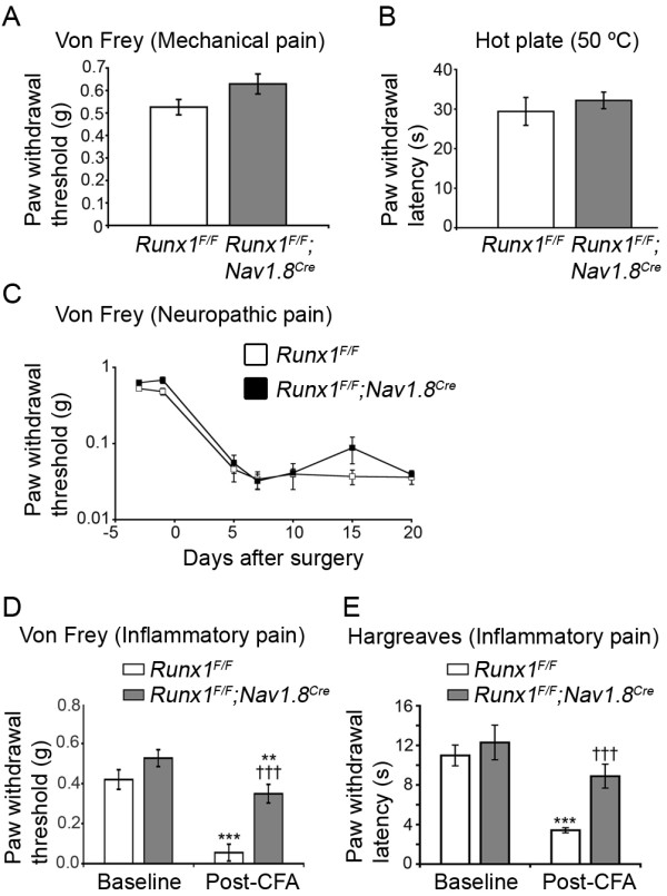
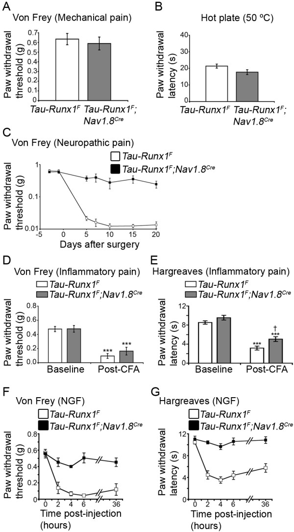
Background: The cellular and molecular programs that control specific types of pain are poorly understood. We reported previously that the runt domain transcription factor Runx1 is initially expressed in most nociceptors and controls sensory neuron phenotypes necessary for inflammatory and neuropathic pain.

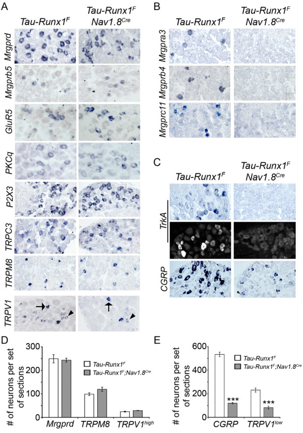
Results: Here we show that expression of Runx1-dependent ion channels and receptors is distributed into two nociceptor populations that are distinguished by persistent or transient Runx1 expression. Conditional mutation of Runx1 at perinatal stages leads to preferential impairment of Runx1-persistent nociceptors and a selective defect in inflammatory pain. Conversely, constitutive Runx1 expression in Runx1-transient nociceptors leads to an impairment of Runx1-transient nociceptors and a selective deficit in neuropathic pain. Notably, the subdivision of Runx1-persistent and Runx1-transient nociceptors does not follow the classical nociceptor subdivision into IB4+ nonpeptidergic and IB4- peptidergic populations.

Conclusion: Altogether, we have uncovered two distinct Runx1-dependent nociceptor differentiation programs that are permissive for inflammatory versus neuropathic pain. These studies lend support to a transcription factor-based distinction of neuronal classes necessary for inflammatory versus neuropathic pain.


Figures

Figure 1
Figure 1
Loss of a subset of Runx1-dependent genes in Runx1F/F;Nav1.8Cre L-CKO mice. (A) Schematic showing the timing of "early" Runx1 knockout (using Wnt1-Cre mice, removing Runx1 before the onset of Runx1 expression), and late Runx1 knockout (using Nav1.8-Cre, removing Runx1 during E16-E17). (B-D) In situ hybridization (ISH) using the indicated probes for Runx1-dependent channels and receptors on transverse sections through adult lumbar (L4/L5) DRG from Runx1F/F control mice and Runx1F/F;Nav1.8Cre L-CKO mice. For Ret and P2X3, double labeling of mRNA (red) with IB4 (green) is shown. For TRPV1 (D), both ISH (top pannels) and immunohistochemistry (IHC) (bottom panels) data are shown. The average numbers of Ret+ and P2X3+ neurons were decreased by 67%, from 599 ± 35 to 199 ± 10 (p < 0.01), and by 56%, from 630 ± 24 to 276 ± 30 (p < 0.001), respectively. Note that the remaining Ret+ and P2X3+ neurons in mutant mice are IB4-. (E) Graph showing the average (± SEM) of the total number of neurons expressing the indicated probes per set of lumbar DRG sections of control (white bar) and mutant (grey bar) mice. Note that the average number of Mrgprb4+ neurons was not significantly changed: from 35 ± 4 to 29 ± 10 (p > 0.05). The number of TRPV1high neurons (arrows) was reduced by 75%, from 21 ± 3 to 5 ± 1 (p < 0.01), whereas the number of TRPV1low (arrowheads) was unchanged, from 210 ± 22 to 218 ± 10 (p > 0.05).
Figure 2
Figure 2
Expression of Runx1-dependent genes in Runx1-persistent versus Runx1-transient neurons in the adult lumbar DRG. (A) Double staining of Runx1 protein (green) and indicated RNA probe (red) on transverse sections of wild-type P30 lumbar (L5) DRG. Quantitative data are shown to the right of the panels. Note that 44.9% (231/515) and 9.2% (72/785) of Runx1+ neurons coexpressed Mrgprd and TRPM8, respectively. Note also that TRPV1high neurons represent 9% of total TRPV1+ neurons (33/356). Arrows indicate double-labeled neurons and arrowheads indicate single-labeled neurons. For TRPV1, asterisks indicate TRPV1low double-labeled neurons. (B) Schematic depicting the segregation of Runx1-dependent genes in the adult DRG into Program A and Program B, expressed predominantly in Runx1-persistent and Runx1-transient nociceptors, respectively.
Figure 3
Figure 3
Runx1F/F;Nav1.8Cre mice showed impaired inflammatory pain but largely unaffected heat pain or neuropathic pain. (A) Mechanical thresholds measured by Von Frey filaments in control mice (n = 10) and L-CKO mice (n = 12). No difference was observed (p > 0.05). (B) Heat sensitivity measured using the hot plate assay. Controls, n = 10. Mutants, n = 12. No difference was observed (p > 0.05). (C) Neuropathic pain (SNI model). No difference was observed in mechanical allodynia over the examined time course (controls, n = 10; mutants, n = 12) (p > 0.05, ANOVA). (D, E) Inflammatory Pain (CFA model), (D) Measurement of mechanical allodynia using Von Frey filaments. Controls showed an 84 ± 3% drop in mechanical threshold two days after CFA injection (n = 9, ***p < 0.001), while mutants showed only a 33 ± 8% drop (n = 14, **p < 0.01). This difference in mechanical sensitivity drop was highly significant (†††p < 0.001). (E) Measurement of heat hyperalgesia using the Hargreaves apparatus. Controls (n = 8) were strongly sensitized (70 ± 4% drop in latency, ***p < 0.001) while mutants (n = 8) showed no significant sensitization (p > 0.05). The difference in latency between controls and mutants post-CFA was highly significant (†††p < 0.001). Error bars indicate standard errors of the mean (SEM).
Figure 4
Figure 4
Selective loss of Runx1-dependent genes in prospective Runx1-transient nociceptors in Tau-Runx1F;Nav1.8Cre mice. (A-C) In situ hybridization (ISH) using the indicated probes on sections through adult lumbar (L4/L5) DRG of Tau-Runx1F control and Tau-Runx1F;Nav1.8Cre mutant mice. For TrkA (C), both ISH (top pannels) and immunohistochemistry (IHC) (bottom panels) data are shown. Arrows indicate neurons expressing TRPV1high and arrowheads indicate those with TRPV1low. (D, E) Graph showing the average (± SEM) of the total number of neurons expressing the indicated probes per set of lumbar DRG sections of control (white bar) and mutant (gray bar) mice. The numbers of Mrgprd+, TRPM8+ and TRPV1high neurons per set of sections (D) were not significantly changed in mutant versus control animals (249 ± 18 versus 243 ± 8 for Mrgprd+ neurons; 99 ± 6 versus 120 ± 9 for TRPM8+ neurons; and 25 ± 2 versus 29 ± 1 for TRPV1high neurons) (p > 0.05). However, CGRP+ and TRPV1low neurons in the mutants (E) decreased from 533 ± 19 to 120 ± 6 (***p < 0.001) and from 231 ± 18 to 82 ± 12 (***p < 0.001), respectively.
Figure 5
Figure 5
Tau-Runx1F;Nav1.8Cre mice exhibited a defect in neuropathic pain but maintained normal heat and CFA-induced inflammatory pain. (A) Mechanical thresholds measured by Von Frey filaments in controls (n = 16) and mutants (n = 10). No difference was observed (p > 0.05). (B) Heat sensitivity measured using the hot plate assay. No significant difference was observed between controls (n = 16) and mutants (n = 10) (p > 0.05). (C) Neuropathic pain (SNI). Measurement of mechanical allodynia using Von Frey filaments. A significant difference was observed over the examined time course (p < 0.001, ANOVA) between controls (n = 14) and mutants (n = 10). (D-E) Inflammatory pain (CFA), (D) Measurement of mechanical allodynia. Both controls and mutants were strongly sensitized two days after CFA injection (Controls: n = 15; ***p < 0.001. Mutants: n = 11, ***p < 0.001) with no significant difference between the two genotypes post-CFA (p > 0.05). (E) Measurement of heat hyperalgesia two days after CFA injection. Both controls and mutants were sensitized (controls: n = 5, ***p < 0.001. Mutants: n = 5; ***p < 0.001). The degree of sensitization in controls (63 ± 3% reduction in latency) was only slightly higher than that in mutants (49 ± 4%) (p < 0.05). (F-G) Pain hypersensitivity in response to intraplantar NGF is impaired in mutant mice. A significant difference between controls (n = 5) and mutants (n = 4) was observed over the examined time course for both mechanical allodynia (p < 0.00001, ANOVA) (F) and thermal hyperalgesia (p < 0.00001, ANOVA) (G). Error bars indicate standard errors of the mean (SEM).

References

    1. Julius D, Basbaum AI. Molecular mechanisms of nociception. Nature. 2001;413:203–210. doi: 10.1038/35093019. - DOI - PubMed
    1. Woolf CJ, Ma Q. Nociceptors-noxious stimulus detectors. Neuron. 2007;55:353–364. doi: 10.1016/j.neuron.2007.07.016. - DOI - PubMed
    1. Basbaum AI, Bautista DM, Scherrer G, Julius D. Cellular and molecular mechanisms of pain. Cell. 2009;139:267–284. doi: 10.1016/j.cell.2009.09.028. - DOI - PMC - PubMed
    1. Pezet S, McMahon SB. Neurotrophins: mediators and modulators of pain. Annu Rev Neurosci. 2006;29:507–538. doi: 10.1146/annurev.neuro.29.051605.112929. - DOI - PubMed
    1. Hucho T, Levine JD. Signaling pathways in sensitization: toward a nociceptor cell biology. Neuron. 2007;55:365–376. doi: 10.1016/j.neuron.2007.07.008. - DOI - PubMed

Publication types

Substances

LinkOut - more resources