Comparative Study
. 2011 Dec;32(12):2323.e27-40.
doi: 10.1016/j.neurobiolaging.2010.06.010. Epub 2010 Jul 31.

Nuclear localization sequence of FUS and induction of stress granules by ALS mutants

Affiliations
Comparative Study

Nuclear localization sequence of FUS and induction of stress granules by ALS mutants

Jozsef Gal et al. Neurobiol Aging. 2011 Dec.

Abstract

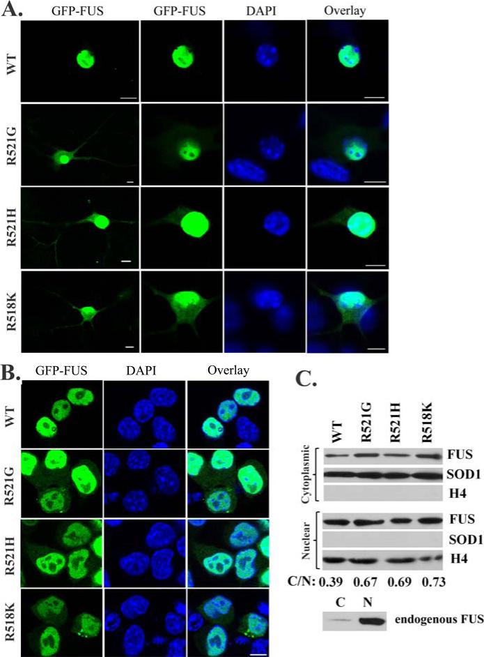
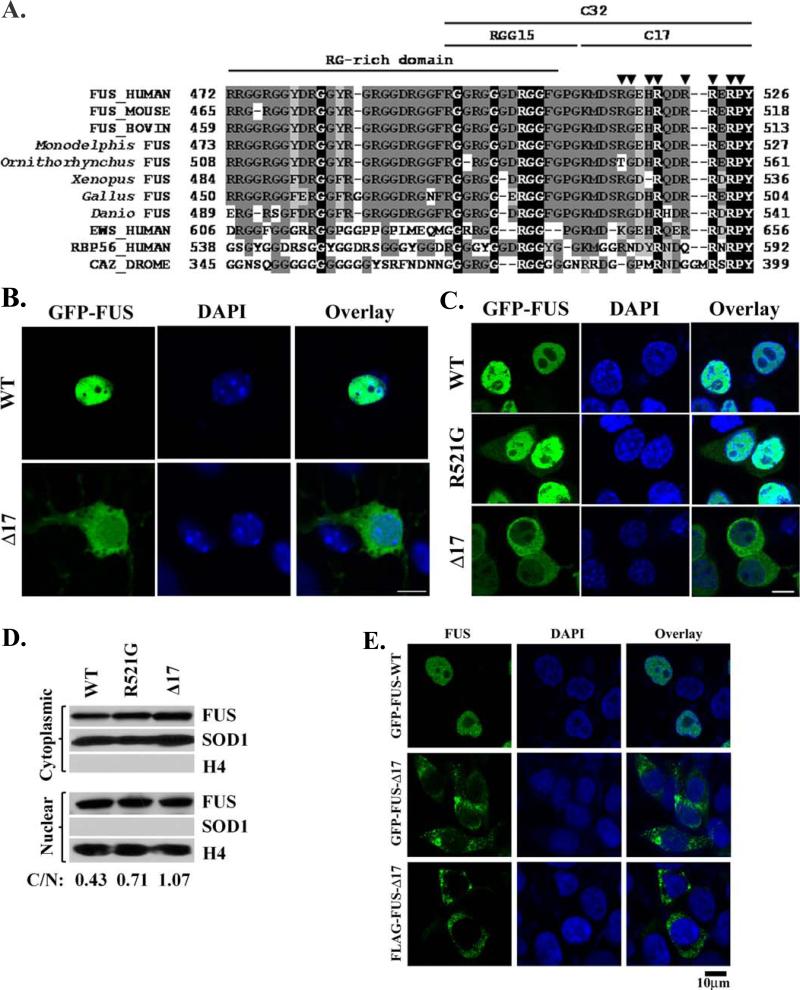
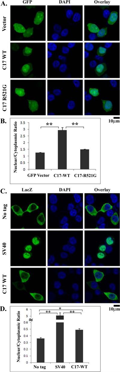
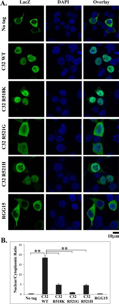
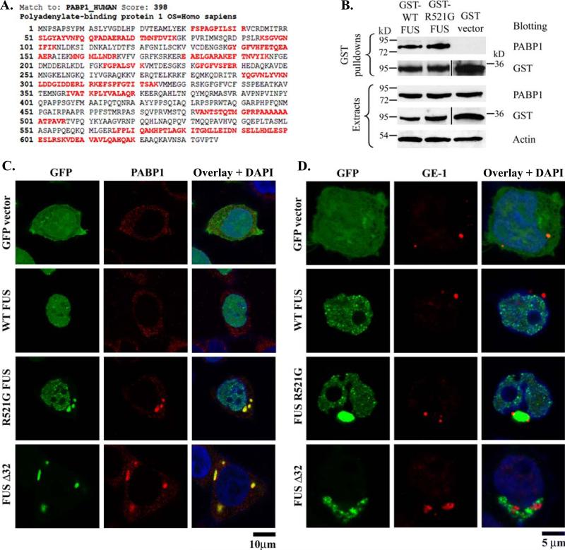
Mutations in fused in sarcoma (FUS) have been reported to cause a subset of familial amyotrophic lateral sclerosis (ALS) cases. Wild-type FUS is mostly localized in the nuclei of neurons, but the ALS mutants are partly mislocalized in the cytoplasm and can form inclusions. We demonstrate that the C-terminal 32 amino acid residues of FUS constitute an effective nuclear localization sequence (NLS) as it targeted beta-galactosidase (LacZ, 116 kDa) to the nucleus. Deletion of or the ALS mutations within the NLS caused cytoplasmic mislocalization of FUS. Moreover, we identified the poly-A binding protein (PABP1), a stress granule marker, as an interacting partner of FUS. Large PABP1-positive cytoplasmic foci (i.e. stress granules) colocalized with the mutant FUS inclusions but were absent in wild-type FUS-expressing cells. Processing bodies, which are functionally related to stress granules, were adjacent to but not colocalized with the mutant FUS inclusions. Our results suggest that the ALS mutations in FUS NLS can impair FUS nuclear localization, induce cytoplasmic inclusions and stress granules, and potentially perturb RNA metabolism.


Figures

Figure 1
Figure 1. Cytoplasmic mislocalization of the familial ALS mutant FUS in primary motor neurons and N2A cells
(A). Confocal microscopic images of mouse embryonic primary motor neurons transfected with GFP-tagged WT or mutant FUS. Partial localization in the cytoplasm was observed for the ALS mutants R521G, R521H and R518K, but WT FUS was mostly localized in the nucleus. (B). Confocal microscopic images of N2A cells transfected with GFP-tagged WT or mutant FUS. Similarly, cytoplasmic localization of mutant FUS was observed. In addition, inclusions were also observed in cells expressing mutant FUS. (C). Western blot of the cytoplasmic and nuclear fractions of cells expressing WT or mutant FUS. Increased levels of mutant FUS were found in the cytoplasmic fraction as compared to WT FUS. The quantification of the relative abundance of the cytoplasmic FUS normalized against the nuclear FUS was shown as C/N values at the bottom of the figure. A nuclear protein histone 4 (H4) and a cytoplasmic protein superoxide dismutase 1 (SOD1) were used to demonstrate the purity of the fractions. Western blot of the endogenous FUS in N2A cells showed that the endogenous WT FUS was largely in the nuclear fraction while a weak band was also found in the cytoplasmic fraction. All scale bars are 10 μm.
Figure 2
Figure 2. The C-terminal 17 residues are essential to the nuclear localization of FUS
(A). The sequence alignment of the C-terminal regions of FUS from multiple organisms, Drosophila CaZ and several human proteins closely related to FUS. X: identical residues; X: strong conservation; X: weaker conservation/similarity. ▼ indicates the positions of known familial ALS mutations. The RG-rich domain, RGG15 motif, C-terminal 17-residue sequence (C17) and C-terminal 32-residue segment (C-32) are marked. (B). Confocal microscopic images of mouse embryonic primary motor neurons transfected with GFP-tagged WT or C17 deletion mutant FUS. FUS-Δ17 was observed in the perikaryon, neurites and nucleus while WT FUS was mostly localized in the nucleus. (C). Similar cytoplasmic localization of FUS-Δ17 was also observed in N2A cells. In addition, inclusions were also observed in cells expressing mutant FUS. (D). Western blot of the cytoplasmic and nuclear fractions of cells expressing WT FUS, R521G or Δ17 mutants. Increased levels of mutant FUS were found in the cytoplasmic fraction as compared to WT FUS. The quantification of the relative abundance of the cytoplasmic FUS normalized against the nuclear FUS was shown as C/N values at the bottom of the figure. H4 and SOD1 were used as nuclear and cytoplasmic markers, respectively. (E). Similar cytoplasmic localization and inclusions of FUS-Δ17 were also observed in HEK293 cells using GFP-tagged FUS or immunofluorescence staining of FLAG-tagged FUS. All scale bars are 10 μm.
Figure 3
Figure 3. The effect of the C-terminal 17 residues on the nuclear localization of GFP and LacZ
(A). Confocal images of HEK293 cells transfected with GFP vector control, GFP-C17, or GFP-C17-R521G. Tagging the C-terminal 17 residues of FUS to GFP caused significant accumulation of GFP-C17 in the nucleus. The ALS mutation R521G within the C-17 segment caused nearly even distribution of GFP-C17-R521 in the entire cell. (B). The average ratios of nuclear and cytoplasmic fluorescence intensities (N/C) were quantified from at least 20 randomly selected cells, see Materials and Methods. (C). Confocal images of HEK293 cells transfected with LacZ alone (no tag), LacZ-SV40, or LacZ-C17. Tagging LacZ with the NLS of the large T antigen of SV40 caused significant relocalization of LacZ-SV40 to the nucleus. In contrast, LacZ-C17 largely remained in the cytoplasm with relatively weak fluorescence in the nucleus. (D). Quantification of the nuclear and cytoplasmic fluorescence intensities as the N/C ratios. The C-terminal 17 residues caused a mild but statistically significant increase of the N/C ratio of LacZ-C17 as compared to LacZ with no tag. SEM were calculated as error bars and Student's t-test was performed to obtain p values in (B) and (D). *, p < 0.0001; **, p < 10-6. All scale bars are 10 μm.
Figure 4
Figure 4. The C-terminal 32 residues are an effective nuclear localization sequence (NLS)
(A). Confocal images of HEK293 cells transfected with LacZ (no tag), LacZ-C32, LacZ-C32-R521G, LacZ-C32-R518K, LacZ-C32-R521H, or LacZ-RGG15. LacZ-C32 was highly concentrated in the nucleus. The ALS mutations (R521G, R518K or R521H) in the C-32 sequence all increased the cytoplasmic localization of the fusion protein. The R521G mutation caused approximately even distribution of LacZ-C32-R521G between the cytoplasm and nucleus while the cytoplasmic fluorescence intensity of LacZ-C32-R518K or LacZC32-R521H was weaker. (B). Quantification of the nuclear and cytoplasmic fluorescence intensities as the N/C ratios. The C-terminal 32 residues were highly effective in increasing the N/C ratio. All three ALS mutations caused significant decrease in the N/C ratios, among which R521G caused the largest decrease. Tagging the RGG15 motif to LacZ did not induce significant change in the N/C ratio compared to the LacZ with no tag. Errors bars represent SEM and p values were calculated using Student's t-test. **, p < 10-6. All scale bars are 10 μm.
Figure 5
Figure 5. The familial ALS mutant FUS co-localized with stress granules
(A). Identification of polyadenylate-binding protein 1 (PABP-1) in the GST-FUS pull-down sample by LC-MS/MS. The identified tryptic peptides within PABP-1 by tandem mass spectrometry (MS/MS) are shown in red. The MASCOT score of 398 suggests a high-confidence identification. (B). GST vector control, GST-WT-FUS or GST-R521G-FUS was transfected to HEK293 cells. The GST pull-down samples were subjected to PABP-1 Western blot. PABP-1 was co-precipitated with GST-FUS but not with the GST vector control. (C). Confocal images of GFP, PABP-1 immunofluorescence and DAPI were obtained from HEK293 cells transfected with GFP vector control, GFP-WT-FUS, GFP-R521G-FUS, or GFP-FUS-Δ32. PABP-1 showed dispersed puncta throughout the cytoplasm in cells transfected with GFP vector or WT FUS. In the cells expressing the R521G or C32 deletion mutant FUS, PABP-1 formed large cytoplasmic foci that resembled stress granules. The stress granules were co-localized with the mutant FUS inclusions. (D). Confocal images of GFP, GE-1 immunofluorescence and DAPI were obtained from HEK293 cells transfected with GFP vector control, GFP-WT-FUS, GFP-R521G-FUS, or GFP-FUS-Δ32. The processing bodies marker GE-1 was adjacent to, but not co-localized with the cytoplasmic inclusions of mutant FUS.

References

    1. Aman P, Panagopoulos I, Lassen C, Fioretos T, Mencinger M, Toresson H, Hoglund M, Forster A, Rabbitts TH, Ron D, Mandahl N, Mitelman F. Expression patterns of the human sarcoma-associated genes FUS and EWS and the genomic structure of FUS. Genomics. 1996;37(1):1–8. - PubMed
    1. Anderson P, Kedersha N. Stress granules: the Tao of RNA triage. Trends Biochem Sci. 2008;33(3):141–50. - PubMed
    1. Andersson MK, Stahlberg A, Arvidsson Y, Olofsson A, Semb H, Stenman G, Nilsson O, Aman P. The multifunctional FUS, EWS and TAF15 proto-oncoproteins show cell type-specific expression patterns and involvement in cell spreading and stress response. BMC Cell Biol. 2008;9:37. - PMC - PubMed
    1. Arai T, Hasegawa M, Akiyama H, Ikeda K, Nonaka T, Mori H, Mann D, Tsuchiya K, Yoshida M, Hashizume Y, Oda T. TDP-43 is a component of ubiquitin-positive tau-negative inclusions in frontotemporal lobar degeneration and amyotrophic lateral sclerosis. Biochem Biophys Res Commun. 2006;351(3):602–11. - PubMed
    1. Baechtold H, Kuroda M, Sok J, Ron D, Lopez BS, Akhmedov AT. Human 75-kDa DNA-pairing protein is identical to the pro-oncoprotein TLS/FUS and is able to promote D-loop formation. J Biol. Chem. 1999;274(48):34337–42. - PubMed

Publication types

MeSH terms

Substances