

Review
doi: 10.1016/j.cbpa.2010.07.002.
Epub 2010 Jul 31.
Development of antibody surrogates for the treatment of cancers and autoimmune disease
Affiliations
- PMID: 20674470
- PMCID: PMC3050540
- DOI: 10.1016/j.cbpa.2010.07.002
Item in Clipboard
Review
Development of antibody surrogates for the treatment of cancers and autoimmune disease
Curr Opin Chem Biol.
2010 Dec.
Abstract
Some therapeutic monoclonal antibodies function by focusing the killing power of the immune system on particular cellular targets, a process known as antibody-dependent cell-mediated cytotoxicity (ADCC). There is considerable interest in developing small-molecule therapeutics that utilize this mechanism of action and so act as a kind of antibody surrogate. Progress and challenges in this interesting field are reviewed here.
Copyright © 2010 Elsevier Ltd. All rights reserved.
Figures
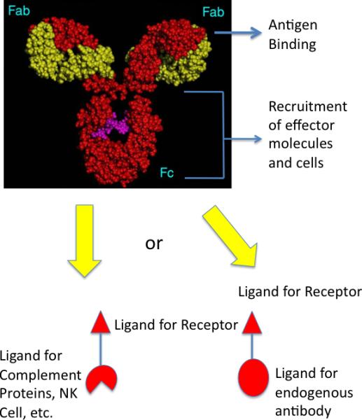
Comparison of a native IgG antibody and a hypothetical synthetic model. Native antibodies have two antigen binding pockets in their Fab regions and a constant region (Fc) capable of interacting with effector molecules and cells, such as the complement proteins, macrophages, natural killer cells, etc. Antibodies could be made by joining a high affinity and selectivity protein-binding molecule to one or more ligands for effector molecules or cells (left). Alternatively, the receptor ligand could be coupled to a molecule bound tightly by an endogenous antibody, whose Fc region would then act to recruit the effector molecules (right; also see Fig. 2)
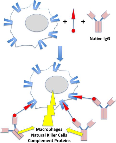
Illustration of ADCC (antibody-dependent cell-mediated cytotoxicity) mediated by a bifunctional molecule capable of binding to a receptor on the surface of the target cell as well as an endogenous IgG antibody. The yellow lightening blot represents the attack of the effector proteins and cells on the target cell. See text for details.

Structures of trapoxen, a naturally occurring HDAC inhibitor, and K-trap, a trapoxen derivative tethered to a solid support. The latter was a key reagent in the identification of HDACs as the target of trapoxen.
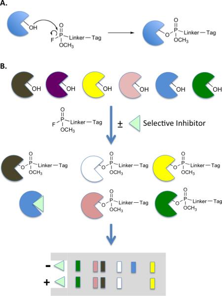
Activity-based protein profiling (ABPP) as a tool for monitoring inhibitor selectivity. A. Fluorophosphonate chemistry for ABPP of serine hydrolases. B. Protocol for testing the selectivity of a serine hydrolase inhibitor (light green triangle). The figure depicts an ideal situation in which six serine hydrolases (different colored pies) are treated with a tagged fluorophosphonate in the presence or absence of the inhibitor. If the inhibitor is specific for the blue hydrolase, then its signal in the profile will disappear, but that of the other proteins will be unaffected. The figure depicts the use of gel-based analysis with blotting specific for the tag to monitor protein labeling.
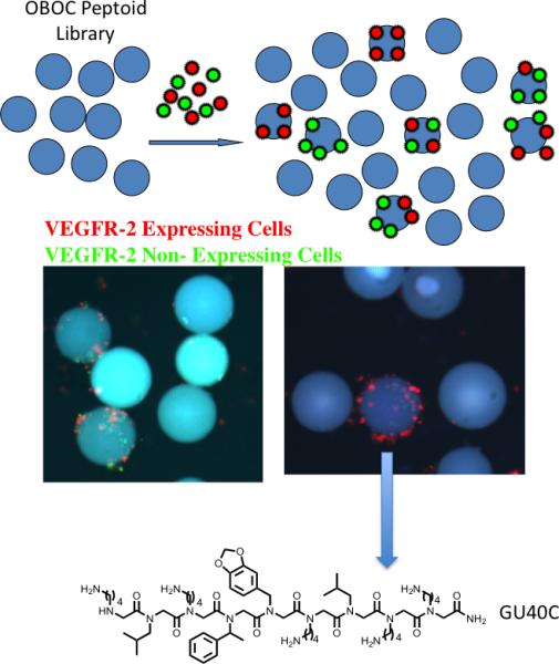
A screening protocol for the identification of highly selective ligands for an integral membrane protein [17]••. See text for details. The fluorescent micrographs show the results from a screen employing cells that do (red-labeled) or do not (green-labeled) express VEGFR2. When an OBOC library of peptoids containing about 260,000 compounds was employed, several thousand beads displaying bound red and green cells were observed (left micrograph). Only five beads that bound almost red cells almost exclusively were observed. The structure of the peptoid, GU40C, displayed by the bead pictured is shown.
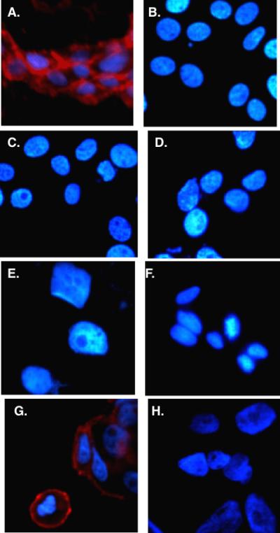
Live cell fluorescence imaging of cells that do (panels A and G) or do not (all other panels) express VEGFR2 incubated with biotinylated GU40C4 (a dimeric version of GU40C (Fig. 5) and red streptavidin-coated quantum dots. The nucleus of the cell was stained blue in each case.
References
-
- Hale G, Clark M, Waldmann H. Therapeutic potential of rat monoclonal antibodies: isotype specificity of antibody-dependent cell-mediated cytotoxicity with human lymphocytes. J Immunol. 1985;134:3056–3061. - PubMed
-
- Matthews R. The B cell slayer. Science. 2007;318:1232–1233. - PubMed
-
- De Palma R, Sementa A. Rituximab in relapsing-remitting multiple sclerosis. N Engl J Med. 2008;358:2645–2646. author reply 2646-2647. - PubMed
-
- Hauser SL, Waubant E, Arnold DL, Vollmer T, Antel J, Fox RJ, Bar-Or A, Panzara M, Sarkar N, Agarwal S, et al. B-cell depletion with rituximab in relapsing-remitting multiple sclerosis. N Engl J Med. 2008;358:676–688. - PubMed
-
- Chaudhuri A, Behan PO. Rituximab in relapsing-remitting multiple sclerosis. N Engl J Med. 2008;358:2646. author reply 2646-2647. - PubMed
Publication types
MeSH terms
Substances
Grants and funding
LinkOut - more resources
Full Text Sources
Other Literature Sources
Medical
