. 2010 Oct 27;170(3):961-70.
doi: 10.1016/j.neuroscience.2010.07.036. Epub 2010 Jul 29.

A transgenic mouse model of neuroepithelial cell specific inducible overexpression of dopamine D1-receptor

Affiliations

A transgenic mouse model of neuroepithelial cell specific inducible overexpression of dopamine D1-receptor

K Fujimoto et al. Neuroscience. .

Abstract

Dopamine and its receptors appear in the brain during early embryonic period suggesting a role for dopamine in brain development. In fact, dopamine receptor imbalance resulting from impaired physiological balance between D1- and D2-receptor activities can perturb brain development and lead to persisting changes in brain structure and function. Dopamine receptor imbalance can be produced experimentally using pharmacological or genetic methods. Pharmacological methods tend to activate or antagonize the receptors in all cell types. In the traditional gene knockout models the receptor imbalance occurs during development and also at maturity. Therefore, assaying the effects of dopamine imbalance on specific cell types (e.g. precursor versus postmitotic cells) or at specific periods of brain development (e.g. pre- or postnatal periods) is not feasible in these models. We describe a novel transgenic mouse model based on the tetracycline dependent inducible gene expression system in which dopamine D1-receptor transgene expression is induced selectively in neuroepithelial cells of the embryonic brain at experimenter-chosen intervals of brain development. In this model, doxycycline-induced expression of the transgene causes significant overexpression of the D1-receptor and significant reductions in the incorporation of the S-phase marker bromodeoxyuridine into neuroepithelial cells of the basal and dorsal telencephalon indicating marked effects on telencephalic neurogenesis. The D1-receptor overexpression occurs at higher levels in the medial ganglionic eminence (MGE) than the lateral ganglionic eminence (LGE) or cerebral wall (CW). Moreover, although the transgene is induced selectively in the neuroepithelium, D1-receptor protein overexpression appears to persist in postmitotic cells. The mouse model can be modified for neuroepithelial cell-specific inducible expression of other transgenes or induction of the D1-receptor transgene in other cells in specific brain regions by crossbreeding the mice with transgenic mouse lines available already.


Figures

Figure 1
Figure 1
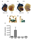
Expression of rtTA in hemizygous nestin-rtTA mouse embryos analyzed by X-gal histochemistry (A – D) and quantitative real-time PCR (E). Expression of the IRES-linked βgeo reporter gene was analyzed using X-gal histochemistry βin whole embryo (A, B) and in coronal sections of the rostral forebrain (C, D). The X-gal reaction product was restricted to the central nervous system in embryonic day 11 (E11) and E13 hemizygous nestin-rtTA embryos and was absent from wild type (WT) littermates. In the rtTA embryos, the reaction product was uniform and intense in the telencephalon (Te), mesencephalon (Me), rhombencephalon (Rh) and the spinal cord (SC). It was absent from skeletal muscle and viscera. Coronal Vibratome sections of the E11 brain (D, E) confirmed robust X-gal labeling in the medial ganglionic eminence (MGE) of the basal forebrain and traces of reaction product in the lateral ganglionic eminence (LGE) and the dorsal cerebral wall (CW). D is a higher magnification view of the labeling. Quantitative real-time PCR of RNA isolated from samples of the MGE, LGE, caudal ganglionic eminence (CGE), CW and striatal differentiating fields (STR) collected from E13 brain sections by laser capture microdissection showed nearly 8-fold higher expression of rtTA mRNA in the MGE compared to the other brain regions confirming the intense X-gal labeling in the MGE.
Figure 2
Figure 2
D1R-EGFP transgene induction in E14 double transgenic (DT) embryos. (A) Transgene mRNA was detected by PCR in forebrain samples obtained from E14 DT mice but not wild type (WT) littermates following doxycycline administration for 2 days, on E12 and E13. (B) Transgene induction was doxycycline dose dependent and occurred at doxycycline doses of 50 or 100 mg/kg/day but not at 0 or 25 mg/kg/day. The transgene induction occurred in forebrain samples of DT embryos and not in WT embryos even though both genotypes were exposed to the same doxycycline dose in utero. (C) Approximately 1,796-fold higher induction of the D1R-EGFP transgene occurred in the DT embryos exposed to doxycycline (arrows) compared to DT embryos not exposed to doxycycline (arrowhead). The extremely low level of expression of the transgene in the DT embryos in the absence of doxycycline (arrowheads) is likely background. DNA base-pair ladder is show in the left lane.
Figure 3
Figure 3
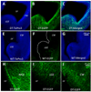
Immunohistochemistry reveals EGFP reporter (surrogate for D1R-EGFP transgene) expression in the forebrain of E14 embryos exposed to doxycycline (50 mg/kg/day) on E12 and E13. EGFP expression (green) is detected in double transgenic (DT; A–C; H–J) embryos but not in wild type (WT; E–G) littermates. Nuclei are labeled with ToPro-3 (blue). EGFP expression occurs in the medial and lateral ganglionic eminences (MGE and LGE, respectively) and in the cerebral wall (CW). Expression in the MGE (H) is more intense than that in the LGE (I) or CW (J). Many EGFP expressing cells resemble neuroepithelial cells in that they have long processes directed away from the lateral ventricle (LV) and towards the pial surface.
Figure 4
Figure 4
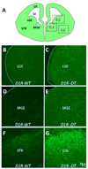
Immunohistochemistry reveals dopamine receptor (D1R) overexpression in the forebrain of E14 wild type (WT) and double transgenic (DT) littermate embryos exposed to doxycycline (50 mg/kg/day) on E12 and E13. A cartoon of a coronal section through an E14 forebrain shows the brain regions in which D1R expression was analyzed (A). D1R expression is significantly higher in the DT embryo compared to the WT embryo in the lateral ganglionic eminence (LGE; B, C), medial ganglionic eminence (MGE, D, E) and striatal differentiating fields (STR, F, G). In both the genotypes, the striatal differentiating fields show the highest D1R expression (F, G) among all the regions analyzed. Scale bar in G = 50µm and applies to all micrographs.
Figure 5
Figure 5
Immunohistochemistry for D1-receptor (D1R) and nestin in the medial ganglionic eminence (MGE) of E14 wild type (WT) and double transgenic (DT) littermate embryos exposed to doxycycline (50 mg/kg/day) on E12 and E13. Lower magnification images show D1R and nestin expression in the MGE of WT (A, B) and DT (C, D) embryos. The D1R labeling in the MGE of the WT (A) is low, compared to that in the DT embryo (C). The higher D1R expression in the MGE of the DT embryo is due to doxycycline-induced overexpression of the D1R tranegene. The higher magnification images of the MGE from the DT embryos show cell surface labeling with the D1R antibody (E) and its co-localization with nestin labeling (F). TUNEL histochemistry (G – J) did not reveal positive profiles in E14 wild type (WT) or DT embryos exposed to doxycycline (50 mg/kg/day) on E12 and E13 in the MGE (G, H), where transgene expression was the highest or in the striatal the differentiating fields (STR; I, J), where the D1R overexpression was the highest. Inset (K) shows an image from a positive control section showing TUNEL+ profiles (green). The control section is from an adult mouse brain subjected to controlled cortical impact from our earlier study (Whalen et al., 2008). Sections processed for TUNEL (E – I) were counterstained with ToPro3 to label all nuclei (blue).
Figure 6
Figure 6
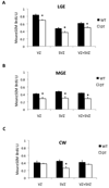
Bromodeoxyuridine (BrdU) labeling index (LI) in the lateral ganglionic eminence (LGE; A), medial ganglionic eminence (MGE; B) and neuroepithelium of the dorsal cerebral wall (CW; C) in embryonic day 14 (E14) double transgenic (DT) and wild type (WT) littermates following doxycycline exposure (50 mg/kg/day) in utero on E12 and E14. The BrdU exposure was for 2 hr. The BrdU LI is significantly decreased in the ventricular zone (VZ), subventricular zone (SVZ) and VZ + SVZ of the LGE and MGE (A, B) and in the SVZ of the CW of the DT embryo compared to the WT littermates.

Similar articles

Cited by

References

    1. Aoki Y, Huang Z, Thomas SS, Bhide PG, Huang I, Moskowitz MA, Reeves SA. Increased susceptibility to ischemia-induced brain damage in transgenic mice overexpressing a dominant negative form of SHP2. Faseb J. 2000;14:1965–1973. - PubMed
    1. Araki KY, Fujimura S, MacDonald ME, Bhide PG. Characterization of mouse striatal precursor cell lines expressing functional dopamine receptors. Dev Neurosci. 2006;28:518–527. - PMC - PubMed
    1. Araki KY, Sims JR, Bhide PG. Dopamine receptor mRNA and protein expression in the mouse corpus striatum and cerebral cortex during pre- and postnatal development. Brain Res. 2007;1156:31–45. - PMC - PubMed
    1. Bhide PG. Cell cycle kinetics in the embryonic mouse corpus striatum. J Comp Neurol. 1996;374:506–522. - PubMed
    1. Bhide PG. Dopamine, cocaine and the development of cerebral cortical cytoarchitecture: A review of current concepts. Seminars Cell Dev Biol. 2009;20:395–402. - PubMed

Publication types