. 2010 Aug 4;12(2):166-73.
doi: 10.1016/j.cmet.2010.05.016.

Identification of surface residues on Niemann-Pick C2 essential for hydrophobic handoff of cholesterol to NPC1 in lysosomes

Affiliations

Identification of surface residues on Niemann-Pick C2 essential for hydrophobic handoff of cholesterol to NPC1 in lysosomes

Michael L Wang et al. Cell Metab. .

Abstract

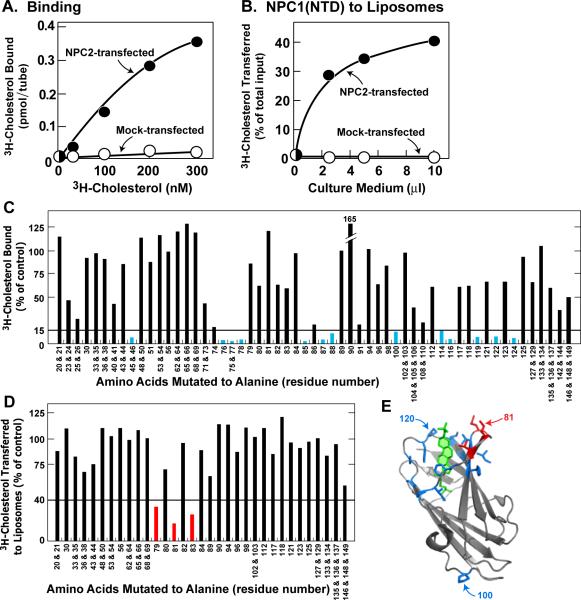
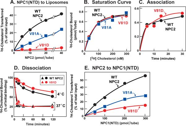
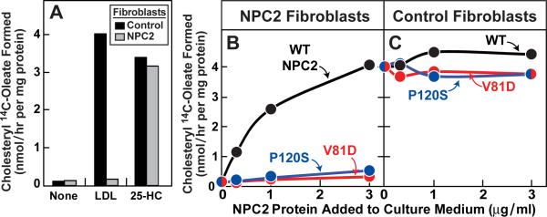
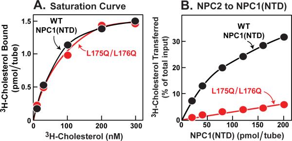
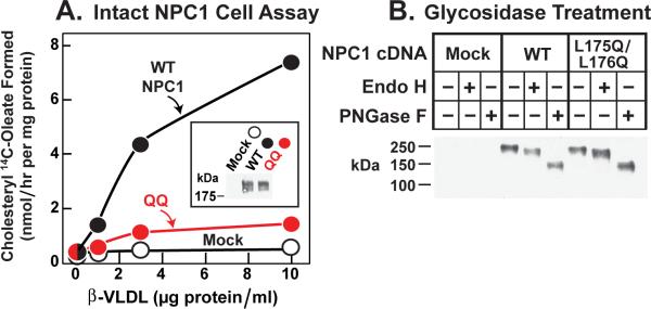
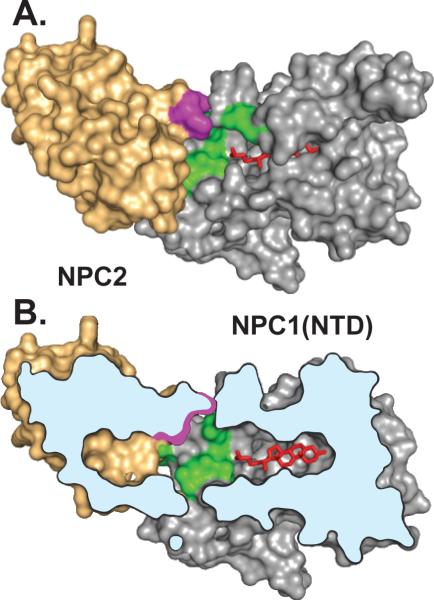
Water-soluble Niemann-Pick C2 (NPC2) and membrane-bound NPC1 are cholesterol-binding lysosomal proteins required for export of lipoprotein-derived cholesterol from lysosomes. The binding site in NPC1 is located in its N-terminal domain (NTD), which projects into the lysosomal lumen. Here we perform alanine-scanning mutagenesis to identify residues in NPC2 that are essential for transfer of cholesterol to NPC1(NTD). Transfer requires three residues that form a patch on the surface of NPC2. We previously identified a patch of residues on the surface of NPC1(NTD) that are required for transfer. We present a model in which these two surface patches on NPC2 and NPC1(NTD) interact, thereby opening an entry pore on NPC1(NTD) and allowing cholesterol to transfer without passing through the water phase. We refer to this transfer as a hydrophobic handoff and hypothesize that this handoff is essential for cholesterol export from lysosomes.


Figures

Figure 1
Figure 1. Alanine Scan Mutagenesis of NPC2
(A and C) [3H]Cholesterol binding to NPC2 in culture medium from transfected cells. Each reaction, in final volume of 160 μl, contained 110 μl buffer B, 50 μl concentrated medium as described in Experimental Procedures, 1.9 μg BSA, 0.004% NP-40, and [3H]cholesterol (A, indicated concentration at 222×103 dpm/pmol; C, 200 nM at 132×103 dpm/pmol). After 2 hr at 4°C, the amount of bound [3H]cholesterol was measured as described in Experimental Procedures except reactions were not diluted before loading onto Ni-NTA columns. (A) Each value represents total binding after subtraction of blank value (<0.03 pmol/tube). (C) Each bar (average of duplicate assays; mean variation ± SEM for all duplicate values, 9.6 ± 1.2%) represents binding relative to WT NPC2 studied in same experiment. All binding data were adjusted for variations (typically <2-fold) in the amount of secreted NPC2 protein as determined by densitometric scanning of immunoblots of the assayed protein in the eluate. The data in the graph were obtained in 5 separate experiments. The “100% of control” values (WT NPC2) averaged 1.5 pmol/tube (average value for mock-transfected cells in same 5 experiments was <0.1 pmol/tube). The entire alanine scan was repeated in an independent experiment with similar results. Blue bars denote residues that when mutated to alanine decrease binding by >85%. (B and D) [3H]Cholesterol transfer from NPC1(NTD) to liposomes in presence of NPC2 contained in culture medium from transfected cells. Each reaction, in a final volume of 200 μl, contained 130 μl buffer A (pH 5.5), ~40 pmol of WT NPC1(NTD)-LVPRGS-His8-FLAG complexed to [3H]cholesterol (132×103 dpm/pmol), 20 μg PC liposomes, and concentrated medium (B, 0–10 μl; D, 30 μl). After 10 min at 4°C, [3H]cholesterol transferred to liposomes was measured as described in Experimental Procedures (assay C). (B) Each value represents percentage of [3H]cholesterol transferred after subtraction of percentage in absence of medium (11% transfer). The 100% value for transfer was 0.14 pmol/tube. (D) Each bar (average of duplicate assays; mean variation ± SEM for all duplicate values, 8.8 ± 1.1%) denotes percentage of [3H]cholesterol transferred in presence of NPC2 after subtraction of percentage in absence of NPC2. The data in the graph were obtained in 5 separate experiments. The “100% of control” values (WT NPC2) averaged 38% transfer of total input (average value for mock-transfected cells in same 5 experiments was 1.8%). The entire alanine scan was repeated in an independent experiment with similar results. Red bars denote residues that when mutated to alanine decrease NPC2-mediated [3H]cholesterol transfer by >60%. (E) Ribbon diagram of bovine NPC2 (Xu et al., 2007), showing positions of residues crucial for cholesterol binding (blue) and transfer (red). Residue P100 is only one of 14 residues important for binding [3H]cholesterol that does not map to sterol-binding pocket. Residue 81 (Ile bovine sequence; Val in human) was identified in above alanine scan as essential for [3H]cholesterol transfer, but not binding. Residue P120 was previously identified as essential for both binding and transfer (Infante et al., 2008c). Bound cholesterol sulfate is shown in green. (C–E) Amino acid residues are numbered starting at the initiator methionine (residue no. 1). The first amino acid after signal peptide cleavage is residue no. 20.
Figure 2
Figure 2. Biochemical Analysis of NPC2 Transfer-defective Mutants
(A) [3H]Cholesterol transfer from NPC1(NTD) to liposomes as a function of NPC2. Each reaction, in a final volume of 200 μl buffer A (pH 5.5), contained ~50 pmol of WT NPC1(NTD)-LVPRGS-His8-FLAG complexed to [3H]cholesterol (222×103 dpm/pmol), 20 μg PC liposomes, and the indicated concentration of WT or mutant NPC2-His10. After 10 min at 4°C, [3H]cholesterol transferred to liposomes was measured as described in Experimental Procedures (assay C). Each value represents percentage of [3H]cholesterol transferred. A blank value in the absence of NPC2 (7% transfer) was subtracted. The 100% value for transfer was 1.9 pmol/tube. (B) [3H]Cholesterol binding. Each reaction, in a final volume of 80 μl buffer A (pH 5.5) with 0.004% NP-40, contained 8 pmol of purified WT or mutant NPC2-His10, 1 μg BSA, and the indicated concentration of [3H]cholesterol (222×103 dpm/pmol). After 2 hr at 4°C, bound [3H]cholesterol was measured. Each value represents total binding after subtraction of blank value (<0.1 pmol/tube). (C) Time course of association of [3H]cholesterol to NPC2. Each reaction, in a final volume of 80 μl of buffer A (pH 5.5) with 0.004% NP-40, contained 8 pmol of WT or mutant NPC2-His10 and 200 nM [3H]cholesterol (132×103 dpm/pmol). After incubation for indicated time at 4°C, bound [3H]cholesterol was determined. Each value represents total binding after subtraction of a blank value (<0.03 pmol/tube). (D) Dissociation of previously bound [3H]cholesterol from NPC2 at different temperatures. Dissociation of [3H]cholesterol from [3H]cholesterol:NPC2-His10 (WT or mutant) was measured as described in Experimental Procedures. Each value represents the percentage of [3H]cholesterol remaining bound to WT or mutant NPC2 relative to zero-time value. The “100% initial binding” values at zero time for NPC2 was 0.48 (WT) and 0.26 (mutant) pmol/tube. (E) [3H]Cholesterol transfer from NPC2 to NPC1(NTD). Each reaction, in a final volume of 200 μl buffer A (pH 5.5), contained ~40 pmol of WT or mutant NPC2-His10 complexed to [3H]cholesterol (222×103 dpm/pmol) and the indicated concentration of WT NPC1(NTD)-LVPRGS-His8-FLAG. After 10 min at 4°C, [3H]cholesterol transferred was measured as described in Experimental Procedures (assay B). Each value represents percentage of [3H]cholesterol transferred to NPC1(NTD). Blank values in the absence of NPC1(NTD) (0.1–0.5% transfer) were subtracted. The 100% values for transfer from WT, V81A, and V81D NPC2 were 0.24, 0.78, and 0.29 pmol/tube, respectively (A–E) Each value is the average of duplicate assays.
Figure 3
Figure 3. Ability of WT NPC2, but not Mutant NPC2, to Rescue LDL-stimulated Cholesteryl Ester Formation in NPC2-deficient Human Fibroblasts
(A) Control and NPC2-deficient cells were set up for experiments as described in Experimental Procedures. On day 7, cells were switched to medium B containing 5% lipoprotein-deficient serum, 50 μM compactin, and 50 μM sodium mevalonate in the absence or presence of 10 μg/ml 25-hydroxycholesterol (25-HC) or 60 μg protein/ml LDL as indicated. (B and C) On day 7, cells were switched to above medium supplemented with 60 μg protein/ml LDL and indicated concentration of purified WT or mutant NPC2-His10. (A–C) After 5 hr at 37°C, each cell monolayer was pulse-labeled for 1 h with 0.2 mM sodium [14C]oleate (3480 dpm/nmol), and cellular content of cholesteryl [14C]oleate and [14C]triglycerides were determined. Each value is the average of duplicate incubations. Content of [14C]triglycerides in NPC2-deficient fibroblasts treated with 60 μg/ml LDL and 3 μg/ml of WT, V81D, or P120S NPC2-His10 proteins was 3.1, 3.0, and 4.0 nmol/hr per mg protein, respectively.
Figure 4
Figure 4. Biochemical Analysis of NPC1(NTD) Transfer-defective Mutant
(A) [3H]Cholesterol binding. Each reaction, in a final volume of 80 μ1 buffer A (pH 5.5) with 0.004% NP-40, contained 4 pmol purified WT or mutant NPC1(NTD)-LVPRGS-His8-FLAG, 1 μg BSA, and indicated concentration of [3H]cholesterol (132×103 dpm/pmol). After 24 hr at 4°C, bound [3H]cholesterol was measured. Each value represents total binding after subtraction of a blank value (<0.06 pmol/tube). (B) [3H]Cholesterol transfer from NPC2 to NPC1(NTD). Each reaction, in a final volume of 100 μl buffer A (pH 5.5), contained ~24 pmol of NPC2-FLAG complexed to [3H]cholesterol (132×103 dpm/pmol) and indicated concentration of WT or mutant NPC1(NTD)-LVPRGS-His8-FLAG. After 10 min at 4°C, [3H]cholesterol transferred was measured as described in Experimental Procedures (assay A). Each value represents percentage of [3H]cholesterol transferred to NPC1(NTD). A blank value in the absence of NPC1(NTD) (0.3% transfer) was not subtracted. The 100% value for transfer from NPC2 was 0.85 pmol/tube. (A and B) Each value is the average of duplicate assays.
Figure 5
Figure 5. Failure of Mutant L175Q/L176Q Version of Full-length NPC1 to Rescue Cholesteryl Ester Formation in NPC1-defective Hamster Cells
(A) Cholesterol esterification in response to ß-VLDL. NPC1-deficient CHO 4-4-19 cells were transfected on day 1 with 2 μg pcDNA3.1 (mock), WT pCMV-NPC1-His8-FLAG, or its mutant version (L175Q/L176Q) as described in Experimental Procedures. Five hr after transfection, the medium was switched to medium A containing 5% newborn calf lipoprotein-deficient serum. On day 2, the medium was switched to same medium containing 5 μM compactin and 50 μM sodium mevalonate. After 16 hr, fresh medium containing 50 μM compactin, 50 μM sodium mevalonate, and the indicated concentration of ß-VLDL was added. After 5 hr at 37°C, each monolayer was pulse-labeled for 1 hr with 0.2 mM sodium [14C]oleate (7433 dpm/nmol). The cells were then harvested for measurement of their content of cholesteryl [14C]oleate and [14C]triglycerides as described in Experimental Procedures. Each value is the average of duplicate incubations. The content of [14C]triglycerides for mock, WT, and mutant NPC1 transfected cells incubated with 10 μg/ml ß-VLDL was 201, 222, and 129 nmol/hr per mg protein, respectively. Inset shows an immunoblot of whole cell extracts from the various transfected cells probed with anti-FLAG antibody as described in Experimental Procedures. (B) Deglycosidase treatment. NPC1-deficient CHO cells were transfected with the indicated plasmid as in (A). Five hr after transfection, the medium was switched to medium A with 5% FCS. Two days later, cells were harvested, and their solubilized membranes were treated with glycosidase EndoH or PNGaseF and then subjected to immunoblot analysis with anti-FLAG antibody as described in Experimental Procedures. (A and B) Filters were exposed to film for ~10 sec at room temperature.
Figure 6
Figure 6. Conceptual Model Illustrating One Possible Mechanism of Interaction Between NPC2 and NPC1(NTD)
This model is based on the published structures of sterol-bound NPC2 (Xu et al., 2007) and sterol-bound NPC1(NTD) in the putative open conformation (Kwon et al., 2009). A stable complex between NPC2 and NPC1(NTD) has not been demonstrated experimentally, and the interaction models in A and B are hypothetical. (A) Surface representation showing residues in NPC2 (purple) and NPC1(NTD) (green) that are crucial for transfer of cholesterol (red) between NPC2 and NPC1(NTD). The cholesterol molecule is shown after its transfer to NPC1(NTD). The two proteins are positioned so that the openings in their respective sterol-binding pockets are juxtaposed and the planes of the cholesterol-binding pockets in NPC2 and NPC1(NTD) are aligned. (B) Cutaway view of the NPC2:NPC1(NTD) complex shown in A, revealing alignment of the cholesterol-binding pockets and juxtaposition of the surface patches on both proteins postulated to be crucial for protein-protein interaction.

References

    1. Anderson KS. Fundamental mechanisms of substrate channeling. Methods Enzymol. 1999;308:111–145. - PubMed
    1. Babalola JO, Wendeler M, Breiden B, Arenz C, Schwarzmann G, Locatelli-Hoops S, Sandhoff K. Development of an assay for the intermembrane transfer of cholesterol by Niemann-Pick C2 protein. Biol. Chem. 2007;388:617–626. - PubMed
    1. Brown MS, Goldstein JL. A receptor-mediated pathway for cholesterol homeostasis. Science. 1986;232:34–47. - PubMed
    1. Carstea ED, Morris JA, Coleman KG, Loftus SK, Zhang D, Cummings C, Gu J, Rosenfeld MA, Pavan WJ, Krizman DB, Nagle J, Polymeropoulos MH, Sturley SL, Ioannou YA, Higgins ME, Comly M, Cooney A, Brown A, Kaneski CR, Blanchette-Mackie J, Dwyer NK, Neufeld EB, Chang T-Y, Liscum L, Strauss JF, III, Ohno K, Zeigler M, Carmi R, Sokol J, Markie D, O'Neill RR, van Diggelen OP, Elleder M, Patterson MC, Brady RO, Vanier MT, Pentchev PG, Tagle DA. Niemann-Pick C1 disease gene: Homology to mediators of cholesterol homeostasis. Science. 1997;277:228–231. - PubMed
    1. Chikh K, Vey S, Simonot C, Vanier MT, Millat G. Niemann-Pick type C disease: importance of N-glycosylation sites for function and cellular location of the NPC2 protein. Mol. Gen. Metabol. 2004;83:220–230. - PubMed

Publication types

MeSH terms