

Acidic and basic fibroblast growth factors involved in cardiac angiogenesis following infarction
- PMID: 20674996
- PMCID: PMC3061206
- DOI: 10.1016/j.ijcard.2010.07.024
Acidic and basic fibroblast growth factors involved in cardiac angiogenesis following infarction
Abstract
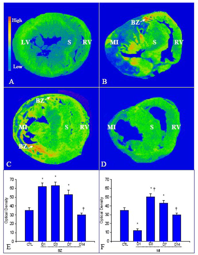
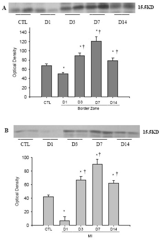
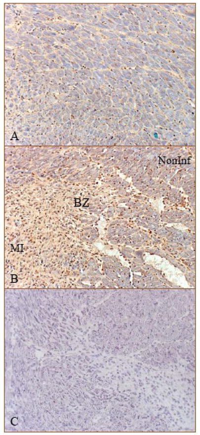
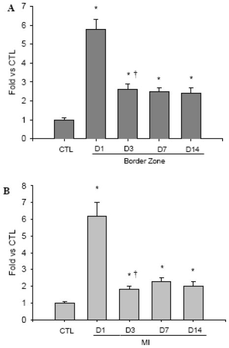
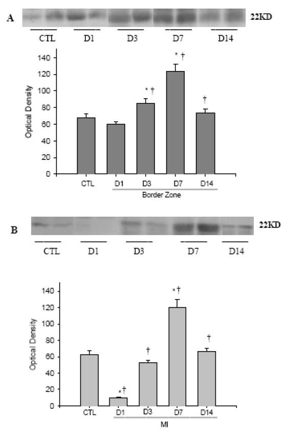
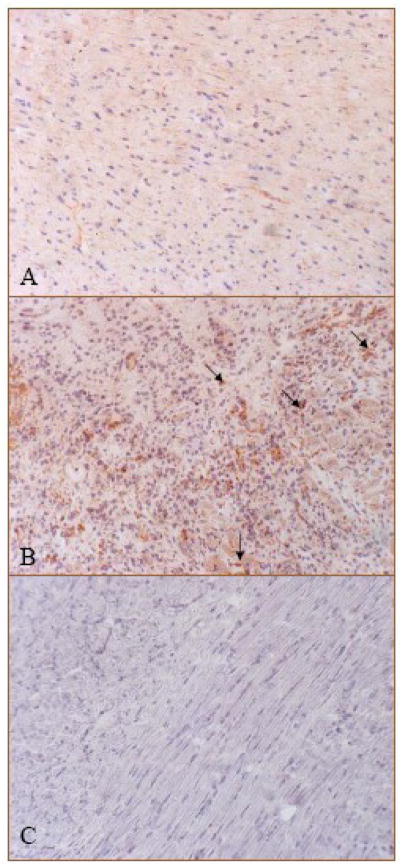
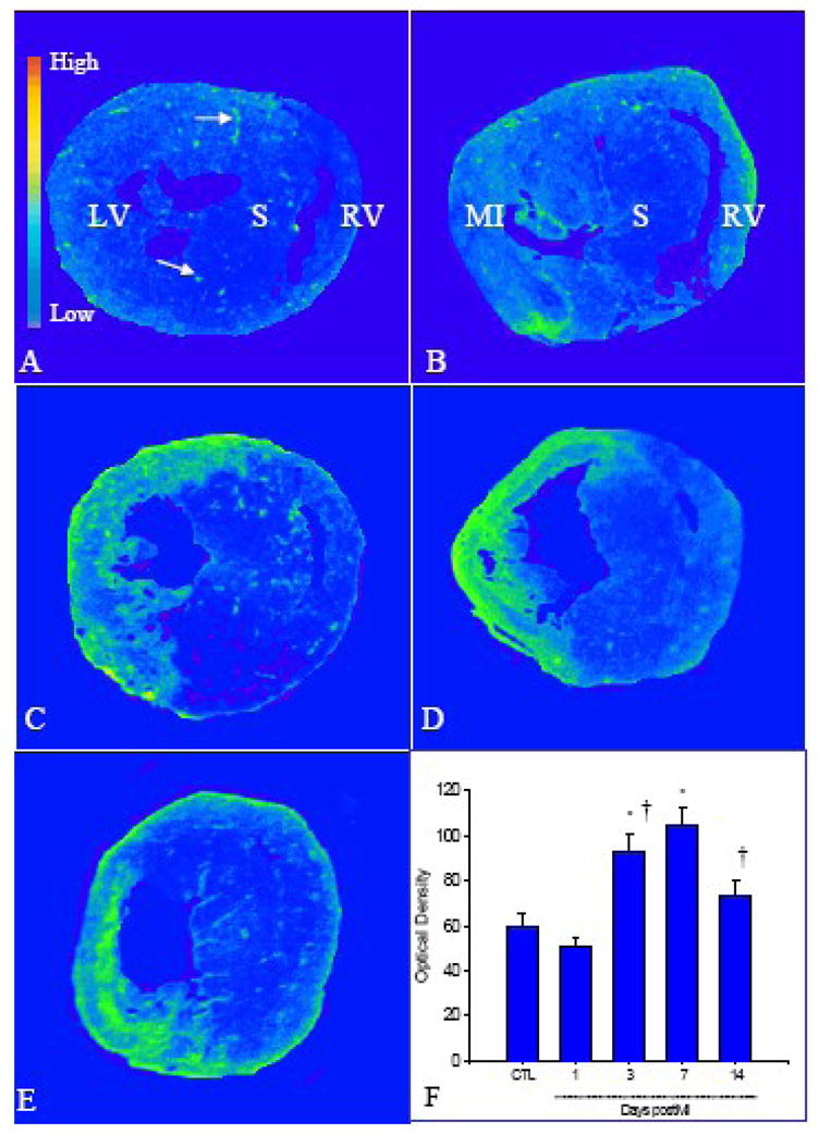
Acidic and basic fibroblast growth factors (FGF-1/FGF-2) promote angiogenesis in cancer. Angiogenesis is integral to cardiac repair following myocardial infarction (MI). The potential regulation of FGF-1/FGF-2 in cardiac angiogenesis postMI remains unexplored. Herein, we examined the temporal and spatial expression of FGF-1/FGF-2 and FGF receptors (FGFR) in the infarcted rat heart at days 1, 3, 7, and 14 postMI. FGF-1/-2 gene and protein expression, cells expressing FGF-1/-2 and FGFR expression were examined by quantitative in situ hybridization, RT-PCR; western blot, immunohistochemistry and quantitative in vitro autoradiography. Compared to the normal heart, we found that in the border zone and infarcted myocardium 1) FGF-1 gene expression was increased in the first week postMI and returned to control levels at week 2; FGF-1 protein levels were, however, largely reduced at day 1, then elevated at day 3 peaked at day 7 and declined at day 14; and cells expressing FGF-1 were primarily inflammatory cells; 2) FGF-2 gene expression was significantly elevated from day 1 to day 14; the increase in FGF-2 protein level was most evident at day 7 and cells expressing FGF-2 were primarily endothelial cells; 3) FGFR expression started to increase at day 3 and remained elevated thereafter; and 4) FGF-1/FGF-2 and FGFR expression remained unchanged in the noninfarcted myocardium. Thus, FGF-1/FGF-2 and FGFR expression are enhanced in the infarcted myocardium in the early stage after MI, which is spatially and temporally coincident with angiogenesis, suggesting that FGF-1/FGF-2 are involved in regulating cardiac angiogenesis and repair.
Copyright © 2010 Elsevier Ireland Ltd. All rights reserved.
Figures
Similar articles
-
Platelet-derived growth factor involvement in myocardial remodeling following infarction.J Mol Cell Cardiol. 2011 Nov;51(5):830-8. doi: 10.1016/j.yjmcc.2011.06.023. Epub 2011 Jul 13. J Mol Cell Cardiol. 2011. PMID: 21767547 Free PMC article.
-
Vascular endothelial growth factor (VEGF)-A: role on cardiac angiogenesis following myocardial infarction.Microvasc Res. 2010 Sep;80(2):188-94. doi: 10.1016/j.mvr.2010.03.014. Epub 2010 Apr 1. Microvasc Res. 2010. PMID: 20362592 Free PMC article.
-
Reactive oxygen species promote angiogenesis in the infarcted rat heart.Int J Exp Pathol. 2009 Dec;90(6):621-9. doi: 10.1111/j.1365-2613.2009.00682.x. Epub 2009 Sep 15. Int J Exp Pathol. 2009. PMID: 19758416 Free PMC article.
-
Molecular mechanisms of angiogenesis: fibroblast growth factor signal transduction.FASEB J. 1995 Jul;9(10):919-25. doi: 10.1096/fasebj.9.10.7542215. FASEB J. 1995. PMID: 7542215 Review.
-
Fibroblast growth factors: at the heart of angiogenesis.Cell Biol Int. 1995 May;19(5):431-44. doi: 10.1006/cbir.1995.1087. Cell Biol Int. 1995. PMID: 7543787 Review.
Cited by
-
VEGF-C/VEGFR-3 pathway promotes myocyte hypertrophy and survival in the infarcted myocardium.Am J Transl Res. 2015 Apr 15;7(4):697-709. eCollection 2015. Am J Transl Res. 2015. PMID: 26064438 Free PMC article.
-
Platelet-derived growth factor blockade on cardiac remodeling following infarction.Mol Cell Biochem. 2014 Dec;397(1-2):295-304. doi: 10.1007/s11010-014-2197-x. Epub 2014 Aug 23. Mol Cell Biochem. 2014. PMID: 25148874
-
Angiogenic growth factors in myocardial infarction: a critical appraisal.Heart Fail Rev. 2017 Nov;22(6):665-683. doi: 10.1007/s10741-017-9630-7. Heart Fail Rev. 2017. PMID: 28639006 Review.
-
Myoblasts and embryonic stem cells differentially engraft in a mouse model of genetic dilated cardiomyopathy.Mol Ther. 2013 May;21(5):1064-75. doi: 10.1038/mt.2013.15. Epub 2013 Feb 26. Mol Ther. 2013. PMID: 23439500 Free PMC article.
-
Platelet-derived growth factor involvement in myocardial remodeling following infarction.J Mol Cell Cardiol. 2011 Nov;51(5):830-8. doi: 10.1016/j.yjmcc.2011.06.023. Epub 2011 Jul 13. J Mol Cell Cardiol. 2011. PMID: 21767547 Free PMC article.
References
-
- Timar J, Dome B, Fazekas K, Janovics A, Paku S. Angiogenesis-dependent diseases and angiogenesis therapy. Pathol Oncol Res. 2001;7:85–94. - PubMed
-
- Ren G, Dewald O, Frangogiannis NG. Inflammatory mechanisms in myocardial infarction. Curr Drug Targets Inflamm Allergy. 2003;2:242–56. - PubMed
-
- Kang DH, Kanellis J, Hugo C, Truong L, Anderson S, Kerjaschki D, et al. Role of the microvascular endothelium in progressive renal disease. J Am Soc Nephrol. 2002;13:806–16. - PubMed
-
- Wheatley-Price P, Shepherd FA. Targeting angiogenesis in the treatment of lung cancer. J Thorac Oncol. 2008;3:1173–84. - PubMed
-
- Roccaro AM, Russo F, Cirulli T, Di Pietro G, Vacca A, Dammacco F. Antiangiogenesis for rheumatoid arthritis. Curr Drug Targets Inflamm Allergy. 2005;4:27–30. - PubMed
Publication types
MeSH terms
Substances
Grants and funding
LinkOut - more resources
Full Text Sources
Other Literature Sources
Medical
