. 2010 Nov;53(5):918-26.
doi: 10.1016/j.jhep.2010.05.015. Epub 2010 Jul 17.

Phosphatidylinositol-3-kinase p110γ contributes to bile salt-induced apoptosis in primary rat hepatocytes and human hepatoma cells

Affiliations

Phosphatidylinositol-3-kinase p110γ contributes to bile salt-induced apoptosis in primary rat hepatocytes and human hepatoma cells

Simon Hohenester et al. J Hepatol. 2010 Nov.

Abstract

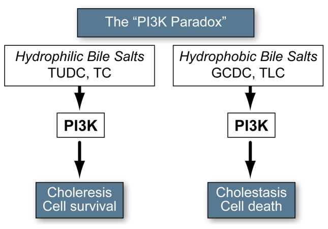
Background & aims: Glycochenodeoxycholate (GCDC) and taurolithocholate (TLC) are hepatotoxic and cholestatic bile salts, whereas tauroursodeoxycholate (TUDC) is cytoprotective and anticholestatic. Yet they all act, in part, through phosphatidylinositol-3-kinase(PI3K)-dependent mechanisms ("PI3K-paradox"). Hepatocytes express three catalytic PI3K Class I isoforms (p110α/β/γ), specific functions of which, in liver, are unclear. In other cell types, p110γ is associated with detrimental effects. To shed light on the PI3K enigma, we determined whether hydrophobic and hydrophilic bile salts differentially activate distinct p110 isoforms in hepatocytes, and whether p110γ mediates bile salt-induced hepatocyte cell death.

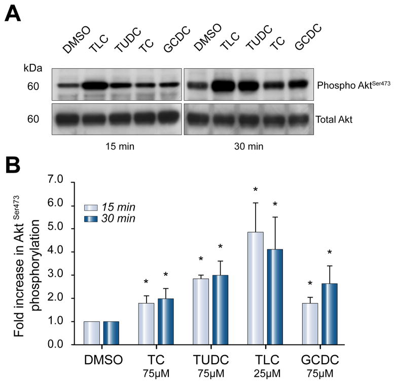
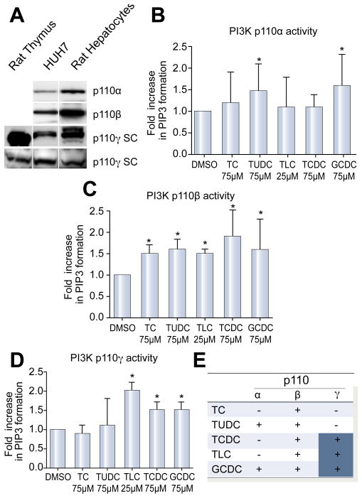
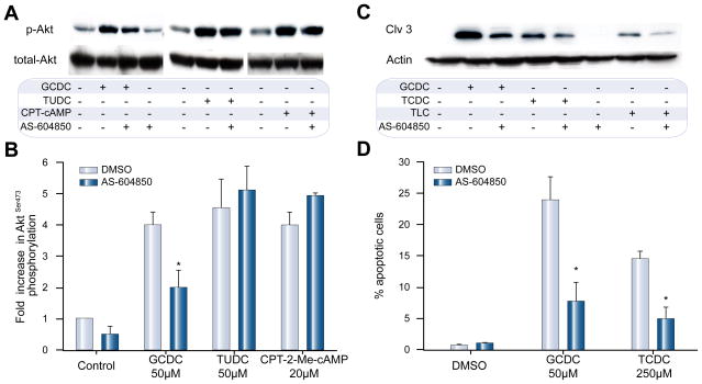
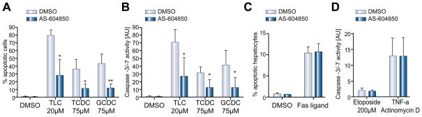
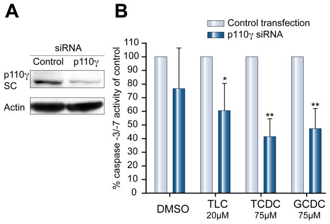
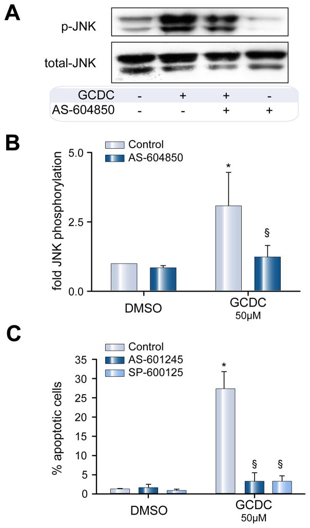
Methods: Isoform-specific PI3K activity assays were established to determine isoform activation by bile salts in rat hepatocytes. Activation of Akt and JNK was determined by immunoblotting. Following stimulation with hydrophobic bile salts, hepatocellular apoptosis was determined morphologically after Hoechst staining and by analysis of caspase-3/-7 activity or caspase-3 cleavage. Activity or expression of PI3K p110γ was inhibited pharmacologically (AS604850) or by knock-down using specific siRNA.

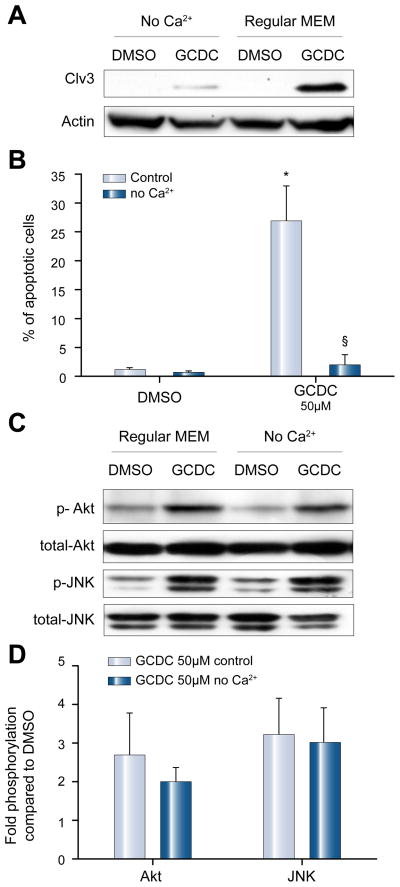
Results: All bile salts tested activated p110β, while p110α was activated by TUDC and GCDC. Intriguingly, only hydrophobic bile salts activated p110γ. Inhibition of p110γ attenuated GCDC-induced Akt- and JNK-activation, but did not alter TUDC- or cAMP-induced Akt-signaling in rat hepatocytes. Inhibition or knock-down of p110γ markedly attenuated hydrophobic bile salt-induced apoptosis in rat hepatocytes and human hepatoma cell lines but did not alter Fas-, tumor necrosis factor α- and etoposide-induced apoptosis. Depletion of Ca(++) prevented GCDC-induced toxicity in rat hepatocytes but did not affect GCDC-induced Akt- and JNK-activation.

Conclusions: PI3K p110γ is activated by hydrophobic, but not hydrophilic bile salts. Bile salt-induced hepatocyte apoptosis is partly mediated via a PI3K p110γ dependent signaling pathway, potentially involving JNK.


Figures

Fig. 1
Fig. 1. Paradoxical role of PI3K in bile salt signaling
PI3K seems to mediate the opposing effects of both hydrophilic and hydrophobic bile salts, promoting both cytotoxic and cytoprotective effects. For additional information see Supplementary Table 1.
Fig. 2
Fig. 2. Hydrophilic and hydrophobic bile salts stimulate Akt-phosphorylation in hepatocytes
Akt phosphorylation levels were determined in lysates of cultured rat hepatocytes by immunoblotting, after stimulation with 25 μM TLC or 50 μM TC, TUDC or GCDC for 15 and 30 min. Equal loading was confirmed by immunoblotting for total-Akt. (A) Representative immunoblots. (B) Densitometric analysis, expressed as fold increase in phosphorylation compared to cells stimulated with DMSO (*p <0.05 vs. DMSO, n = 4).
Fig. 3
Fig. 3. Expression and activation of PI3K p110 isoforms by bile salts in hepatocytes
(A) Expression of p110α, β, and γ in rat hepatocytes and Huh7-Ntcp cells was confirmed by immunoblotting. Two different antibodies (Cell Signaling Technologies(C):CS and Santa Cruz(C):SC) were used for p110γ. Rat thymus served as a positive control for p110γ. Rat hepatocyte cultures were stimulated with bile salts at 25 μM (TLC) to 50 μM (TC, TUDCA, GCDC, TCDC) for 15 min, and isoforms-specific lipid kinase assays were performed. Lipid kinase assays after immunoprecipitation of (B) p110α, (C) p110β, and (D) p110γ (mean ± SD, *p <0.05 vs. DMSO, n = 4–6) are shown. Results represent fold increase in formation of the specific PI3K reaction product PIP3 compared to DMSO. (E) Summary of kinase assays: “+” indicates activation of the respective isoform.
Fig. 4
Fig. 4. Inhibitor of p110γ, AS604850, diminishes GCDC-induced PI3K activation and apoptosis but does not alter TUDC- and CPT-2-Me-cAMP induced signaling in hepatocytes
Rat hepatocytes were stimulated with 50 μM GCDC or TUDC, or 20 μM CPT-2-Me-cAMP, an activator of p110α and p110β, with and without pre-incubation with 2.5 μM AS604850. (A) Akt-phosphorylation was determined by immunoblotting for phosphorylated AktSer-473 and total Akt (representative blots) (B) Densitometric analysis of 3 independent experiments (*p <0.05 vs. control). (C) Rat hepatocytes were pre-treated with AS604850 and then exposed to DMSO, 50 μM GCDC or 250 μM TCDC for 2–4 h, respectively, and apoptosis determined by immunoblotting for the 17 kd cleavage product of active caspase-3 or (D) following staining with Hoechst 33258 (*p <0.05 vs. control, n = 3). Equal loading was confirmed by immunoblotting for actin on the same membrane.
Fig. 5
Fig. 5. Inhibition of p110γ selectively inhibits bile salt-induced apoptosis in hepatocytes but does not alter the effects of independent apoptotic stimuli
HepG2-Ntcp cells were stimulated with DMSO, 20 μM TLC or 75 μM of TCDC or GCDC. The p110γ inhibitor AS604850 (2.5 μM) was added 30 min prior to stimulation. (A) Apoptotic cells were quantified morphologically after Hoechst staining. Rate of apoptosis was expressed as % of apoptotic cells. (B) In separate experiments, apoptosis in identically stimulated HepG2-Ntcp was determined by caspase-3/-7 activity (*p <0.05 vs. control, n = 3–6). (C) Rat hepatocyte cultures were treated with 50 ng/ml Fas ligand for 4 hours and apoptotic cells were determined morphologically. AS604850 was added at 2.5 μM 30 min prior to the addition of Fas ligand (n = 3). (D) HepG2-Ntcp cells were treated with 200 μM etoposide or 200 ng/ml TNFα plus 28 ng/ml actinomycin D for 4 h and apoptotic effects were quantified in caspase-3/-7 assays. Prior to stimulation, cells were treated with 2.5 μM AS604850 for 30 min (n = 3).
Fig. 6
Fig. 6. p110γ knockdown inhibits hydrophobic bile acid-induced increases in caspase-3/-7 activity
(A) Transfection with p110γ siRNA effectively silenced protein expression in Huh7-Ntcp as shown by immunoblots from control transfected cells and cells transfected with specific p110γ siRNA. (B) p110γ-knockdown cells were stimulated with DMSO, 20 μM TLC or 75 μM TCDC or GCDC. After 4 h, activity of caspases-3/-7 was determined and the data are expressed as % of caspase-3/-7 activity of control transfected cells (n = 4, *p <0.05/**p <0.005 vs. control).
Fig. 7
Fig. 7. GCDC-induced apoptosis involves PI3K p110γ-dependent activation of JNK
Rat hepatocytes were pretreated for 30 min with 2.5 μM of the p110γ inhibitor AS604850 and then exposed to 50 μM GCDC for 30 min after which the amount of phosphorylated JNK was determined by immunoblotting. (A) Representative blot and (B) Densitometric analysis of 4 independent experiments (*p <0.05 vs. DMSO, §p <0.05 vs. GCDC control ). (C) Rat hepatocytes were pretreated with either of 2 JNK inhibitors, SP600125 or AS-612245 (10 μM, each) for 30 min and then exposed to 50 μM GCDC for 2 h. Apoptosis was determined morphologically and expressed as % apoptotic cells (n = 3, *p <0.05 vs. DMSO, §p <0.05 vs. GCDC control).
Fig. 8
Fig. 8. GCDC-induced apoptosis but not Akt- and JNK-signaling is calcium dependent
Rat hepatocytes were cultured in Ca++ free medium for 2 h, stimulated with 50 μM GCDC for 2 h and apoptosis was determined by (A) immunoblotting for the active cleavage fragment of caspase-3, and by (B) morphologic evaluation of Hoechst stained cells (n = 4, *p <0.05 vs. DMSO, §p <0.05 vs. control). (C) Rat hepatocytes were cultured in Ca++ free media for 2 h and then exposed to GCDC for 30 min. The amount of phosphorylated Akt and JNK was determined by immunoblotting. Representative immunoblots are shown in (C) and the results of densitometric analysis representing 3 separate experiments are shown in (D). Equal loading was controlled by immunoblotting for total-JNK or total-Akt.

References

    1. Guicciardi ME, Gores GJ. Bile acid-mediated hepatocyte apoptosis and cholestatic liver disease. Dig Liver Dis. 2002;34:387–392. - PubMed
    1. Rust C, Bauchmuller K, Fickert P, Fuchsbichler A, Beuers U. Phosphatidylinositol 3-kinase-dependent signaling modulates taurochenodeoxycholic acid-induced liver injury and cholestasis in perfused rat livers. Am J Physiol Gastrointest Liver Physiol. 2005;289:G88–94. - PubMed
    1. Perez MJ, Briz O. Bile-acid-induced cell injury and protection. World J Gastroenterol. 2009;15:1677–1689. - PMC - PubMed
    1. Wimmer R, Hohenester S, Pusl T, Denk GU, Rust C, Beuers U. Tauroursodeoxycholic acid exerts anticholestatic effects by a cooperative cPKC alpha-/PKA-dependent mechanism in rat liver. Gut. 2008;57:1448–1454. - PubMed
    1. Hohenester S, Oude-Elferink RP, Beuers U. Primary biliary cirrhosis. Semin Immunopathol. 2009 - PMC - PubMed

Publication types

MeSH terms

Substances