. 2011 Jan 1;89(1):157-65.
doi: 10.1093/cvr/cvq251. Epub 2010 Jul 29.

Expansion of human cardiac stem cells in physiological oxygen improves cell production efficiency and potency for myocardial repair

Affiliations

Expansion of human cardiac stem cells in physiological oxygen improves cell production efficiency and potency for myocardial repair

Tao-Sheng Li et al. Cardiovasc Res. .

Abstract

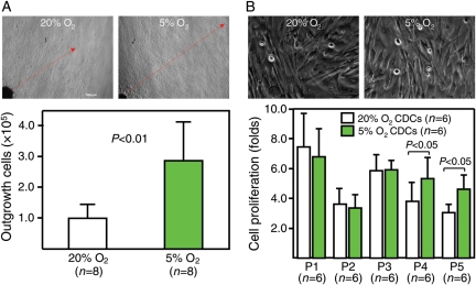
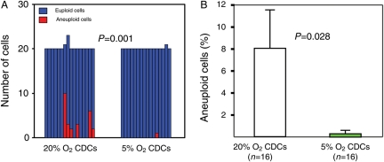
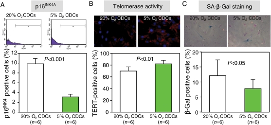
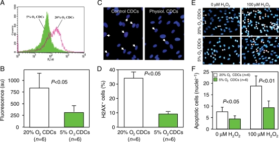
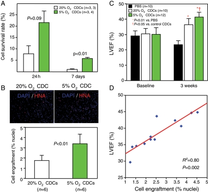
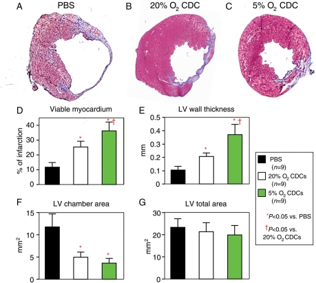
Aims: the ex vivo expansion of cardiac stem cells from minimally invasive human heart biopsies yields tens of millions of cells within 3-4 weeks, but chromosomal abnormalities were frequently detected in preliminary production runs. Here we attempt to avoid aneuploidy and improve cell quality by expanding human cardiac stem cells in physiological low-oxygen (5% O(2)) conditions, rather than in traditional culture in a general CO(2) incubator (20% O(2)).

Methods and results: human heart biopsies (n = 16) were divided and processed in parallel to expand cardiac stem cells under 5% or 20% O(2). Compared with 20% O(2), 5% O(2) culture doubled the cell production and markedly diminished the frequency of aneuploidy. Cells expanded in 5% O(2) showed lower intracellular levels of reactive oxygen species, less cell senescence, and higher resistance to oxidative stress than those grown in 20% O(2), although the expression of stem cell antigens and adhesion molecules was comparable between groups, as was the paracrine secretion of growth factors into conditioned media. In vivo, the implantation of 5% O(2) cells into infarcted hearts of mice resulted in greater cell engraftment and better functional recovery than with conventionally cultured cells.

Conclusion: the expansion of human adult cardiac stem cells in low oxygen increased cell yield, and the resulting cells were superior by various key in vitro and in vivo metrics of cell quality. Physiological oxygen tensions in culture facilitate the ex vivo expansion of healthy, biologically potent stem cells.


Figures

Figure 1
Figure 1
Growth and proliferation of cardiac stem cells in 5% O2 and 20% O2. (A) Representative images (upper) show that cell outgrowth from ‘explants’ (red arrow) was much faster in 5% O2 than in 20% O2. The number of cells harvested (lower bar graph) was more than two-fold higher in 5% O2 than in 20% O2, although the amount of starting material was equivalent. (B) CDCs at earlier passages show no differences in either morphology (upper images, passage #2) or proliferative activity (lower bar graph) under 5% O2 and 20% O2, although greater proliferation was observed in cells expanded under 5% O2 at later passages.
Figure 2
Figure 2
Chromosomal abnormalities. (A) Lower numbers of aneuploid cells were found in CDCs expanded in 5% O2 than in 20% O2. (B) The percentages of aneuploid cells were also decreased in 5% O2 culture.
Figure 3
Figure 3
Cell senescence. Compared with 20% O2 culture, cell senescence of CDCs was improved under 5% O2 culture, by flow cytometry for p16INK4A (A), immunostaining for telomerase activity (B), and senescence-associated β-galactosidase staining (C).
Figure 4
Figure 4
Intracellular ROS, DNA damage, and resistance to oxidative stress. (A) The levels of intracellular ROS are lower in CDCs expanded in 5% O2 when compared with those in 20% O2. (B) DNA damage evidenced by the formation of γ-H2AX foci (upper images) was lower in CDCs expanded in 5% O2 than in 20% O2 (lower bar graph). (C) Representative images (upper images) show TUNEL-positive (red) CDCs after 24 h exposure to 100 µM H2O2. The number of apoptotic cells (lower bar graph) was lower in CDCs expanded in 5% O2 than 20% O2.
Figure 5
Figure 5
Cell engraftment, cardiac functional recovery, and their relationships. (A) Quantitative data on the survival rate of human CDCs 24 h and 7 days after implantation into mice-infarcted heart. (B) Cells positively stained by HNA are more frequently observed in mice 3 weeks after implantation with CDCs expanded in 5% O2 than in 20% O2 (upper images). Lower bar graph shows quantitative data for cell engraftment (% nuclei) in the infarcted heart. (C) LVEF at baseline does not differ among groups, indicating a similar infarct size to begin with. After 3 weeks, the LVEF was higher in mice implanted with CDCs expanded in 5% O2 than in 20% O2, although the LVEF was also higher in mice implanted with CDCs expanded in 20% O2 than in controls with PBS injection only. (D) The engraftment of human CDCs (% nuclei) within the infarcted hearts of mice is strongly correlated with the absolute values of LVEF at 3 weeks.
Figure 6
Figure 6
Histological assessments of infarct size and ventricular morphology. Representative images of Masson's staining show that, compared with an infarcted heart receiving PBS injection only (A), the infarct size was much smaller in a heart that had received CDCs expanded in both 5% O2 (C) and 20% O2 (B). Quantitative analyses of viable myocardium (D), LV wall thickness (E), LV chamber area (F), and LV total area (G) show that better therapeutic efficiency was achieved by the implantation of CDCs expanded in 5% O2 than in 20% O2, although a significant improvement was also observed by the implantation of CDCs expanded under 20% O2 when compared with the control treatment with PBS injection only.

References

    1. Maitra A, Arking DE, Shivapurkar N, Ikeda M, Stastny V, Kassauei K, et al. Genomic alterations in cultured human embryonic stem cells. Nat Genet. 2005;37:1099–1103. doi:10.1038/ng1631. - DOI - PubMed
    1. Baker DE, Harrison NJ, Maltby E, Smith K, Moore HD, Shaw PJ, et al. Adaptation to culture of human embryonic stem cells and oncogenesis in vivo. Nat Biotechnol. 2007;25:207–215. doi:10.1038/nbt1285. - DOI - PubMed
    1. Sareen D, McMillan E, Ebert AD, Shelley BC, Johnson JA, Meisner LF, et al. Chromosome 7 and 19 trisomy in cultured human neural progenitor cells. PLoS ONE. 2009;4:e7630. doi:10.1371/journal.pone.0007630. - DOI - PMC - PubMed
    1. Rubio D, Garcia-Castro J, Martín MC, de la Fuente R, Cigudosa JC, Lloyd AC, et al. Spontaneous human adult stem cell transformation. Cancer Res. 2005;65:3035–3039. - PubMed
    1. Furlani D, Li W, Pittermann E, Klopsch C, Wang L, Knopp A, et al. A transformed cell population derived from cultured mesenchymal stem cells has no functional effect after transplantation into the injured heart. Cell Transplant. 2009;18:319–331. doi:10.3727/096368909788534906. - DOI - PubMed

Publication types

MeSH terms