Review
. 2010 Sep;16(9):1692-724.
doi: 10.1261/rna.2237610. Epub 2010 Jul 30.

Novel endoribonucleases as central players in various pathways of eukaryotic RNA metabolism

Affiliations
Review

Novel endoribonucleases as central players in various pathways of eukaryotic RNA metabolism

Rafal Tomecki et al. RNA. 2010 Sep.

Abstract

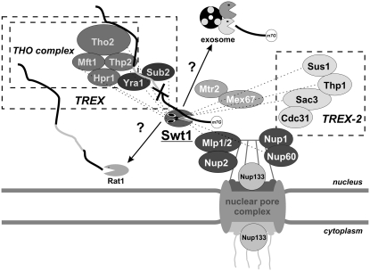
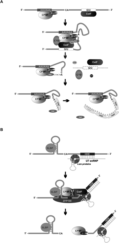
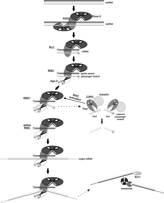
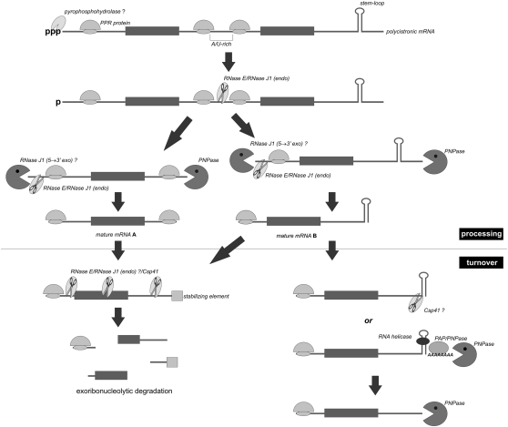
For a long time it has been assumed that the decay of RNA in eukaryotes is mainly carried out by exoribonucleases, which is in contrast to bacteria, where endoribonucleases are well documented to initiate RNA degradation. In recent years, several as yet unknown endonucleases have been described, which has changed our view on eukaryotic RNA metabolism. Most importantly, it was shown that the primary eukaryotic 3' --> 5' exonuclease, the exosome complex has the ability to endonucleolytically cleave its physiological RNA substrates, and novel endonucleases involved in both nuclear and cytoplasmic RNA surveillance pathways were discovered concurrently. In addition, endoribonucleases responsible for long-known processing steps in the maturation pathways of various RNA classes were recently identified. Moreover, one of the most intensely studied RNA decay pathways--RNAi--is controlled and stimulated by the action of different endonucleases. Furthermore, endoribonucleolytic cleavages executed by various enzymes are also the hallmark of RNA degradation and processing in plant chloroplasts. Finally, multiple context-specific endoribonucleases control qualitative and/or quantitative changes of selected transcripts under particular conditions in different eukaryotic organisms. The aim of this review is to discuss the impact of all of these discoveries on our current understanding of eukaryotic RNA metabolism.


Figures

FIGURE 1.
FIGURE 1.
Overview of general RNA metabolism processes in eukaryotic cells involving the action of endoribonucleases and the intracellular localization of the enzymes described in the text. Endonucleolytic cleavages underlie numerous processing events of different RNA classes (rRNA, tRNA, mRNA, snRNA, and snoRNA) occurring in the nuclear compartment, both in the nucleoplasm and the nucleolus. The endoribonucleolytic activity of the exosome Dis3 catalytic subunit participates in the degradation of RNA species such as by-products of rRNA processing and CUTs, while the Swt1 endonuclease is a constituent of RNA surveillance machinery. In the cytoplasm, endonucleases are engaged in RNA quality control pathways dependent on ongoing translation, in the regular turnover of mRNA and in the RNA interference phenomenon. Moreover, Nob1 endonuclease is responsible for the final step of 18S rRNA processing which takes place in the cytoplasm. Finally, in plant chloroplasts, several endoribonucleases are principal enzymes involved in the processing and degradation of organellar transcripts.
FIGURE 2.
FIGURE 2.
Possible model of cooperation between exoribonucleolytic and endoribonucleolytic activities of the exosome complex Dis3 catalytic subunit. (A) Structural viewpoint: RNA substrates containing a secondary structure and lacking the 3′ single-stranded extension of sufficient length are unable to reach the Dis3 exoribonuclease active site via the central channel pathway and are partially resistant to exoribonucleolytic degradation. The PIN domain endoribonuclease activity (scissors) may overcome this problem by cleaving within the loop of the hairpin. The resulting upstream cleavage product might be delivered to the RNB catalytic center either through the channel (top), or via an alternative hypothetical path going directly from the PIN domain to the active site of the RNB domain (middle). In parallel, the RNA substrate can be degraded further by the PIN domain endonuclease before accessing RNB exonuclease (bottom). (B) Functional viewpoint: effects of Dis3 RNB exonuclease and PIN endonuclease catalytic mutations. (left) Cooperative action of the two nucleolytic activities in the wild-type leads to the degradation of a given substrate and its decay intermediates arising due to the endonucleolytic cuts. (center) Inactivation of exonuclease activity alone leads to the accumulation of both a full-length substrate and its endo-cleavage intermediates. (right) Inactivation of both exonuclease and endonuclease activities has a synergistic effect on the level of full-length substrates, whereas the cleavage intermediates disappear in the double mutant.
FIGURE 3.
FIGURE 3.
The Swt1 endoribonuclease is a novel component of nuclear RNA surveillance machinery. Defective pre-mRNA (for instance, containing the 5′ splice site mutation, represented by the cross) gets trapped in the export-incompetent mRNP with the Tho/TREX complex. Endonucleolytic cleavage of the aberrant transcript by Swt1, concentrated in the proximity of the NPC, might facilitate its subsequent exoribonucleolytic degradation, most probably executed by the exosome and Rat1 5′ → 3′ exoribonuclease. The genetic interactions detected between Swt1 and different components of the Tho/TREX complex, as well as with subunits of the TREX-2 complex, coupling transcription with mRNP assembly and export, and with the NPC-associated proteins involved in the perinuclear RNA surveillance by the retention of unspliced transcripts, are marked with dashed lines.
FIGURE 4.
FIGURE 4.
Endonucleases participate in translation-dependent RNA quality control pathways in the cytoplasm. (A) Nonsense-mediated decay; stalling of the ribosome on a premature termination codon leads to the recruitment of SMG1 and UPF1 NMD factors as a result of signal received from poly(A) binding proteins (PABP) bound to the distant poly(A) tail (pA) by eRF1/eRF3 translation termination factors. PTC recognition in higher eukaryotes is also mediated by the EJC deposited upstream of the exon–exon junction following the stop codon. Phosphorylation of UPF1 by SMG1 with the assistance of UPF2/UPF3 proteins leads to a change in UPF1 conformation and triggers decay of the PTC-containing transcript via the use of SMG5/SMG7 NMD factors through deadenylation and decapping (which might be deadenylation-independent or stimulated by removal of poly[A] tail) followed by the degradation executed by the exosome and Xrn1, respectively. In metazoans, an alternative NMD pathway, dependent on SMG6 endoribonucleolytic cleavage in the proximity of PTC, can be used. Upstream and downstream cleavage products are degraded by the exosome and Xrn1, respectively. (B) No-go decay; ribosome stalling due to the secondary structure in mRNA recruits Dom34/Hbs1 NGD factors homologous with eRF1/eRF3 proteins. This enables ribosome release and leads to the degradation of aberrant mRNA initiated by endoribonucleolytic cleavage in the vicinity of the structure blocking translation. Whether the cut is exerted by Dom34 or occurs through the recruitment of an unknown endonuclease remains to be investigated. Subsequent exoribonucleolytic degradation proceeds in a similar manner to that of the NMD pathway. (C) 18S nonfunctional rRNA decay; a block in translation can also result from the 18S rRNA mutations in the decoding center of the small ribosomal subunit (indicated with the gray cross). Such defective 18S rRNA molecules are subjected to the quality control pathway involving Dom34/Hbs1 factors, most probably in a manner similar to that of NGD, including endonucleolytically mediated initiation.
FIGURE 5.
FIGURE 5.
CPSF-73: an endoribonuclease critical for the processing of protein-coding transcripts. (A) Poly(A)+ mRNAs. Most protein-coding transcripts undergo coupled cleavage/polyadenylation reactions. The consecutive steps of this process and the roles of individual cis elements in the mRNA sequence and protein factors acting in trans are described in the main text; the 73-kDa CPSF component is an endonuclease belonging to the metallo-β-lactamase family of enzymes, which usually cleave the pre-mRNA after the CA dinucleotide, defining the site of poly(A) tail addition. (B) Metazoan poly(A)- histone mRNAs. These transcripts employ a noncanonical 3′-end processing mechanism dependent on the binding of U7 snRNP, composed of U7 snRNA and the LSm proteins, to the HDE sequence located downstream from the cleavage site. Another element required for histone mRNA processing is the conserved stem-loop upstream of the cleavage site which is bound by the SLBP factor. Cleavage is preceded by an interaction involving SLBP and U7 snRNP, bridged by the zinc-finger protein ZFP100 and utilizing CPSF and possibly also CstF, similarly to the mechanism presented in A. The CPSF-73 subunit is not only responsible for the endonucleolytic cleavage which determines the position of the histone mRNA mature 3′ end, but it might also display 5′ → 3′ exoribonucleolytic activity, allowing for the degradation of the downstream cleavage product.
FIGURE 6.
FIGURE 6.
Ribosomal RNA maturation in yeast comprises several processing events controlled by different endoribonucleases. The primary transcript synthesized by the rDNA unit is cleaved by an Rnt1 RNase III–like double-stranded specific endonuclease at the site B0 within the 3′ ETS, followed by cleavage at the site A0 in the 5′ ETS carried out by an as yet unidentified enzyme. The Utp24 endonuclease is most likely responsible for cleavages at sites A1 and A2, removing 5′ ETS and separating further steps of 18S (left) and 5.8S/25S (right) rRNA processing, respectively. The 20S pre-rRNA is converted into the mature 18S rRNA in the cytoplasm due to the action of Nob1 endonuclease at site D, probably in cooperation with the Pfa1/Prp43 and Ltv1 proteins. Processing intermediates encompassing 5.8S and 25S rRNA are trimmed at the 5′ end due to the endoribonucleolytic activity of RNase MRP and Rat1/Xrn1 exoribonucleases. The 5.8S and 25S rRNA maturation pathways are subsequently separated by endonucleolytic cleavage at site C2 and completed by exoribonucleolytic processing.
FIGURE 7.
FIGURE 7.
Multiple endoribonucleases participate in different phases of RNA interference in eukaryotic cells. Long dsRNA molecules are processed into 21–25-nt siRNAs by the Dicer RNase III-like enzyme, together with its RNA-binding partner R2D2, leading to the formation of the RISC-loading complex (RLC) which is converted into RISC following the recruitment of Ago-2. The latter enzyme cuts the passenger strand in the RNA duplex, being the first substrate for its endoribonucleolytic activity, into 12- and 9-nt fragments. Cleavage of these RNA species by Trax, a catalytic component of the C3PO endoribonuclease, is a prerequisite for RISC activation, which enables recognition of target mRNA molecules. The targeted transcript is then “sliced” by Ago-2 and finally the RNA fragments located upstream of and downstream from the cleavage site are degraded by the exosome and Xrn1.
FIGURE 8.
FIGURE 8.
mRNA processing and turnover in plant chloroplasts are controlled by the concerted action of endo- and exoribonucleases. The event which most likely initiates the processing of polycistronic chloroplast transcripts is the removal of pyrophosphate from the 5′ end, similarly to the case of prokaryotes. This reaction sensitizes the transcript to attack by endoribonucleases such as RNase E and RNase J1, which cleave polycistron at highly accessible single-stranded AU-rich regions. The 5′ and 3′ ends of the pre-mRNAs thus created are subsequently trimmed by endo- and exonucleases until the stable stem-loop structures or sequences bound by PPR motif–containing proteins, defining the positions of mature mRNA termini. The turnover phase of chloroplast transcripts might be initiated by either the combined action of endoribonucleases cleaving within 5′ UTR, coding sequence or 3′ UTR, followed by exoribonucleolytic decay (left branch) and/or through the removal of the 3′ stem-loop which can occur in two ways: 1) endoribonucleolytic cleavage by the Csp41 enzyme, or 2) cooperative destabilization by the RNA helicase supported by repetitive cycles of polyadenylation and exoribonucleolytic degradation (right branch).
FIGURE 9.
FIGURE 9.
The IRE1 endoribonuclease is a central component of mammalian UPR pathways. ER stress in mammalian cells results in the accumulation of misfolded proteins which elicits three major responses. The most immediate UPR effector pathway is activation of PERK/PEK which upon dimerization and autophosphorylation phosphorylates eIF2α and leads to translational shutdown. Moreover, the ATF6 transmembrane protein is converted by proteolysis into an active transcription factor which stimulates the expression of genes encoding ER chaperones via binding to ESRE sites in their promoters (top). If these early control mechanisms are sufficient for restoring ER homeostasis, the UPR is completed. Otherwise, ATF6 also activates the transcription of the XBP1 gene, producing an unspliced mRNA. At the same time, BiP dissociates from the lumenal domains (LD) of IRE1 monomers, which is the prerequisite for their dimerization/oligomerization (top). The latter process occurs over a series of events, including trans-autophosphorylation of kinase domains (KD) (which might be counteracted by the action of two phosphatases, namely, Ptc2 and Dcr2), binding of the adenosine nucleotide cofactor and reorientation of oligomer subunits, all of which eventually lead to the activation of IRE1 endoribonucleolytic sites in KEN domains (middle). The active IRE1 nuclease has several functions in the late effector phase of UPR (bottom). Above all, it catalyzes cytoplasmic splicing of XBP1 mRNA together with tRNA ligase, thus enabling its translation into an active transcription factor which up-regulates the expression of genes encoding ER chaperones and enzymes degrading unfolded proteins through their ESRE and UPRE promoter elements. On the other hand, IRE1 is also a degradative endonuclease, trigerring the decay of ER-targeted transcripts. The participation of IRE1 in the degradation of 28S rRNA and the autoregulation of its own mRNA levels is also possible, although it is not known yet whether or not IRE1 cleaves them directly (bottom). All of these RNA metabolism events significantly enhance the abilities of cells to efficiently deal with the presence of improperly folded proteins in the ER.
FIGURE 10.
FIGURE 10.
Model of PMR1-mediated cleavage of albumin mRNA. The PMR1 protein is inherently unstable and unless protected by the Hsp90 chaperone it is subjected to the proteasomal degradation pathway. It is maintained in a latent state until the phosphorylation of tyrosine 650, which can be induced, for instance, following the binding of EGF to its receptor. The activated PMR1 is then recruited to its substrate undergoing translation, most likely with the involvement of some unidentified protein partner (designated here as PMR-BP), thereby leading to formation of the polysome-bound PMR1–albumin mRNA complex. Subsequent cleavage of the albumin transcript is followed by dissociation of PMR1 from polysomes.

References

    1. Acharya KR, Shapiro R, Allen SC, Riordan JF, Vallee BL 1994. Crystal structure of human angiogenin reveals the structural basis for its functional divergence from ribonuclease. Proc Natl Acad Sci 91: 2915–2919 - PMC - PubMed
    1. Allmang C, Petfalski E, Podtelejnikov A, Mann M, Tollervey D, Mitchell P 1999. The yeast exosome and human PM–Scl are related complexes of 3′ → 5′ exonucleases. Genes Dev 13: 2148–2158 - PMC - PubMed
    1. Amrani N, Sachs MS, Jacobson A 2006. Early nonsense: mRNA decay solves a translational problem. Nat Rev Mol Cell Biol 7: 415–425 - PubMed
    1. Baillat D, Hakimi MA, Naar AM, Shilatifard A, Cooch N, Shiekhattar R 2005. Integrator, a multiprotein mediator of small nuclear RNA processing, associates with the C-terminal repeat of RNA polymerase II. Cell 123: 265–276 - PubMed
    1. Barnes T, Kim WC, Mantha AK, Kim SE, Izumi T, Mitra S, Lee CH 2009. Identification of Apurinic/apyrimidinic endonuclease 1 (APE1) as the endoribonuclease that cleaves c-myc mRNA. Nucleic Acids Res 37: 3946–3958 - PMC - PubMed

Publication types

LinkOut - more resources