. 2010 Aug 15;24(16):1718-30.
doi: 10.1101/gad.1938710. Epub 2010 Jul 30.

Nf2/Merlin controls progenitor homeostasis and tumorigenesis in the liver

Affiliations

Nf2/Merlin controls progenitor homeostasis and tumorigenesis in the liver

Samira Benhamouche et al. Genes Dev. .

Abstract

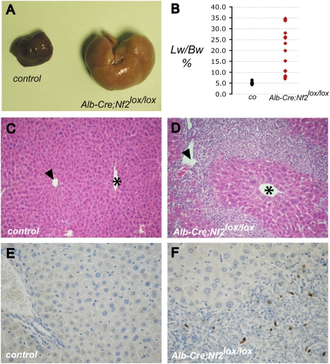
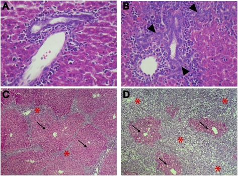
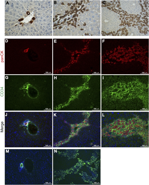
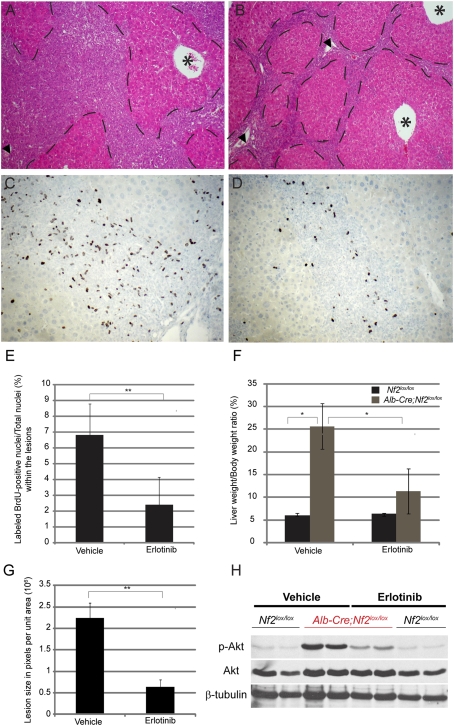
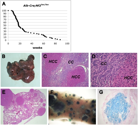
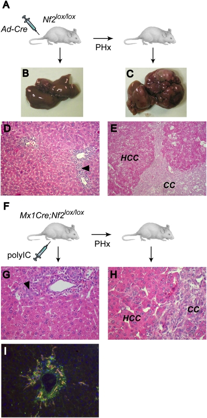
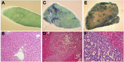
The molecular signals that control the maintenance and activation of liver stem/progenitor cells are poorly understood, and the role of liver progenitor cells in hepatic tumorigenesis is unclear. We report here that liver-specific deletion of the neurofibromatosis type 2 (Nf2) tumor suppressor gene in the developing or adult mouse specifically yields a dramatic, progressive expansion of progenitor cells throughout the liver without affecting differentiated hepatocytes. All surviving mice eventually developed both cholangiocellular and hepatocellular carcinoma, suggesting that Nf2(-/-) progenitors can be a cell of origin for these tumors. Despite the suggested link between Nf2 and the Hpo/Wts/Yki signaling pathway in Drosophila, and recent studies linking the corresponding Mst/Lats/Yap pathway to mammalian liver tumorigenesis, our molecular studies suggest that Merlin is not a major regulator of YAP in liver progenitors, and that the overproliferation of Nf2(-/-) liver progenitors is instead driven by aberrant epidermal growth factor receptor (EGFR) activity. Indeed, pharmacologic inhibition of EGFR blocks the proliferation of Nf2(-/-) liver progenitors in vitro and in vivo, consistent with recent studies indicating that the Nf2-encoded protein Merlin can control the abundance and signaling of membrane receptors such as EGFR. Together, our findings uncover a critical role for Nf2/Merlin in controlling homeostasis of the liver stem cell niche.


Figures

Figure 1.
Figure 1.
Targeted deletion of Nf2 in the mouse liver results in massive hepatic enlargement due to OC hyperproliferation. (A) Hepatomegaly exhibited by the liver of a 20-wk-old Alb-Cre;Nf2lox/lox mouse (right) relative to the liver from a control Nf2lox/lox littermate (left). (B) Distribution of liver weight:body weight ratios in Alb-Cre;Nf2lox/lox (red) versus control (black) mice between 1 and 4 mo of age. Livers of Alb-Cre;Nf2lox/lox mice comprise up to one-third of the body mass. (C,D) Hematoxylin- and eosin-stained paraffin sections of 9-wk-old littermates reveals marked periportal expansion of OCs throughout Alb-Cre;Nf2lox/lox (D) but not control (C) livers. Portal veins (arrowheads) and centrilobular veins (asterisks) are denoted (200×). (E,F) As revealed by BrdU incorporation, proliferation in Alb-Cre;Nf2lox/lox livers is limited to the OCs, and is not seen in surrounding hepatocytes (shown in F). (E) BrdU-incorporating cells are essentially undetectable in the control liver. A single BrdU pulse was given 2 h before sacrifice (400×).
Figure 2.
Figure 2.
Evolution of OC hyperplasia in Alb-Cre;Nf2lox/lox mice. Hematoxylin- and eosin-stained sections reveal that thickening of portal spaces (arrowheads) is already detectable in neonatal (postnatal day 3 [P3]) Alb-Cre;Nf2lox/lox livers (B) compared with Nf2lox/lox control littermate livers (A) (600×). (C,D) Massive and progressive expansion of OCs (asterisks) in the liver of 3-wk-old (C) and 15-wk-old (D) Alb-Cre;Nf2lox/lox mice entraps residual foci of perivenous hepatocytes (arrows) (200×).
Figure 3.
Figure 3.
Expression of classic OC markers in Nf2−/− liver lesions. (A–C) Immunohistochemical analysis using an anti-panCK antibody reveals prominent and progressive staining of the expanding OC population in the liver of 3-wk-old (B) (600×) and 9-wk-old (C) Alb-Cre;Nf2lox/lox mice (200×). (A) In contrast, in the control Nf2lox/lox liver, only the portal bile duct cells express panCK antigens (600×). (D–L) Immunofluorescent detection of panCK and CD34 reveals prominent expression throughout the expanding lesions in 3-wk-old (E,H,K) and 9-wk-old (F,I,L) Alb-Cre;Nf2lox/lox livers, compared with the control Nf2lox/lox liver (D,G,J). (G,J) CD34 staining of portal vein endothelium is also apparent in the control. Costaining of the lesions with anti-CD34 and anti-panCK antibodies is shown in K and L. (M,N) The well-known A6 antibody, which shares epitopes with the panCK antibody (Kofman et al. 2005; Jelnes et al. 2007), similarly exhibits prominent staining throughout the lesions (N), but detects only BECs in the normal liver (M).
Figure 4.
Figure 4.
Deregulated EGFR signaling drives the overproliferation of Nf2−/− liver progenitors in vivo. (A,B) After only 10 d of erlotinib treatment, a reduction of the lesion area (outlined by dashed lies) is apparent in Alb-Cre;Nf2lox/lox mice (B) compared with the Alb-Cre;Nf2lox/lox littermates treated with vehicle 6% captisol (A). Hematoxylin and eosin-stained sections are shown (100×). Portal veins (arrowheads) and centrilobular veins (asterisks) are denoted. (C,D) Immunohistochemical detection of BrdU incorporation reveals a marked decrease in proliferating OCs within the lesions in erlotinib-treated Alb-Cre;Nf2lox/lox mice (D) compared with the vehicle-treated littermates (C) (200×). A single BrdU pulse was given 2 h before sacrifice. (E) Quantification of the erlotinib-induced decrease in BrdU-incorporating OCs in vivo. For each liver sample, ∼3000 to ∼5000 OC nuclei were counted. The graph reflects the number of BrdU-positive nuclei per total nuclei within the lesions across multiple animals (n = 4 for erlotinib group; n = 3 for vehicle group). (**) P < 0.05. (F) Distribution of liver weight:body weight ratios in Nf2lox/lox (gray) versus Alb-Cre;Nf2lox/lox (black) mice treated with vehicle and erlotinib. After only 10 d, erlotinib treatment led to a reduction in the proportion of the total body mass accounted for by the liver (liver weight:body weight ratio), from 25% in vehicle-treated Alb-Cre;Nf2lox/lox mice to 10% in erlotinib-treated Alb-Cre;Nf2lox/lox mice. (*) P < 0.1. (G) Quantification of lesion size in vehicle- and erlotinib-treated mice. Liver lesion size is represented by pixels (occupied by lesion) per total unit area (270 nm2). (**) P < 0.05. (H) In vivo administration of erlotinib yields a decrease in the levels of pAKT in Alb-Cre;Nf2lox/lox livers. Nf2lox/lox mice were used as controls. Western blots of total liver extracts representative of three independent experiments are shown.
Figure 5.
Figure 5.
Metastatic liver tumor development in Alb-Cre;Nf2lox/lox mice. (A) Survival of Alb-Cre;Nf2lox/lox mice. While many animals died within a few months of age, likely due to compromised liver function, those that survived beyond ∼30 wk all developed frank HCC and CC. (B) Multiple tumors are macroscopically evident in a 64-wk-old Alb-Cre;Nf2lox/lox liver. (C,D) Hematoyxlin and eosin staining reveals both CC and HCC neoplasias in the liver of an 83-wk-old Alb-Cre;Nf2lox/lox;GtRosa26lox/stop/lox mouse. (C) 100×. (D) 400×. (E–G) Lung metastases from the liver shown in C. Hematoxylin and eosin staining (40×). HCC metastases in the lung exhibit LacZ expression, as revealed by whole-mount (F) and paraffin section (G).
Figure 6.
Figure 6.
Deletion of Nf2 in the adult liver yields OC hyperproliferation and tumor development that are promoted by PHx. (A) Two-month-old Nf2lox/lox mice were injected with Ad-Cre, followed 2 wk later by two-third PHx or sham operation, and were sacrificed 8 mo to 1 yr post-PHx. The liver from nonhepatectomized mice (B) reveals only mild periportal OC hyperplasia (arrowhead) (D). In contrast, Ad-Cre-injected mice develop multiple liver tumors (C) of both CC and HCC types (E) when subjected to proliferative stimuli induced by PHx. (D,E) Hematoxylin and eosin-stained sections (200×). (F) Two-month-old Mx1-Cre;Nf2lox/lox mice were intraperitoneally injected with polyIC, subjected to PHx or sham operation after 2 wk, and sacrificed at 7–8 mo. (G,H) Hematoxylin and eosin-stained sections reveal mild periportal OC hyperplasia (arrowhead) in control, nonhepatectomized mice (G), and both CC and HCC in mice subjected to PHx (H) (400×). (I) As in Alb-Cre;Nf2lox/lox livers, lesions in hepatectomized, polyIC-induced Mx1-Cre;Nf2lox/lox livers express the OC markers panCK (red) and CD34 (green) (400×).
Figure 7.
Figure 7.
Transplantation of clonal Nf2−/− HBs into the liver of immunocompromised mice yields OC hyperplasia and cholangiocellular tumors. (A,B) Five weeks after transplantation into the liver of nude mice, control HBs are distributed uniformly throughout the liver parenchyma, as indicated by LacZ expression detected by whole-mount (a section of the tissue shown in A is presented in B). (C–F) Five weeks after transplantation, Ad-Cre-infected, Nf2-deficient HBs form multiple foci of undifferentiated LacZ-positive cells (C) that progress to neoplasias (E) exhibiting both hepatocytic and cholangiocytic features (D,F). Hematoxylin and eosin-stained sections are shown in D and F. (B,D) 200×. (F) 400×.

Comment in

References

    1. Adams GB, Chabner KT, Alley IR, Olson DP, Szczepiorkowski ZM, Poznansky MC, Kos CH, Pollak MR, Brown EM, Scadden DT 2006. Stem cell engraftment at the endosteal niche is specified by the calcium-sensing receptor. Nature 439: 599–603 - PubMed
    1. Alison MR 2005. Liver stem cells: Implications for hepatocarcinogenesis. Stem Cell Rev 1: 253–260 - PubMed
    1. Andersen JB, Loi R, Perra A, Factor VM, Ledda-Columbano GM, Columbano A, Thorgeirsson SS 2010. Progenitor-derived hepatocellular carcinoma model in the rat. Hepatology 51: 1401–1409 - PMC - PubMed
    1. Baumgartner R, Poernbacher I, Buser N, Hafen E, Stocker H 2010. The WW domain protein Kibra acts upstream of Hippo in Drosophila. Dev Cell 18: 309–316 - PubMed
    1. Blechacz B, Gores GJ 2008. Cholangiocarcinoma: Advances in pathogenesis, diagnosis, and treatment. Hepatology 48: 308–321 - PMC - PubMed

Publication types

MeSH terms