. 2010 Oct;299(4):L513-22.
doi: 10.1152/ajplung.00274.2009. Epub 2010 Jul 30.

Critical role for lactate dehydrogenase A in aerobic glycolysis that sustains pulmonary microvascular endothelial cell proliferation

Affiliations

Critical role for lactate dehydrogenase A in aerobic glycolysis that sustains pulmonary microvascular endothelial cell proliferation

Glenda Parra-Bonilla et al. Am J Physiol Lung Cell Mol Physiol. 2010 Oct.

Abstract

Pulmonary microvascular endothelial cells possess both highly proliferative and angiogenic capacities, yet it is unclear how these cells sustain the metabolic requirements essential for such growth. Rapidly proliferating cells rely on aerobic glycolysis to sustain growth, which is characterized by glucose consumption, glucose fermentation to lactate, and lactic acidosis, all in the presence of sufficient oxygen concentrations. Lactate dehydrogenase A converts pyruvate to lactate necessary to sustain rapid flux through glycolysis. We therefore tested the hypothesis that pulmonary microvascular endothelial cells express lactate dehydrogenase A necessary to utilize aerobic glycolysis and support their growth. Pulmonary microvascular endothelial cell (PMVEC) growth curves were conducted over a 7-day period. PMVECs consumed glucose, converted glucose into lactate, and acidified the media. Restricting extracellular glucose abolished the lactic acidosis and reduced PMVEC growth, as did replacing glucose with galactose. In contrast, slow-growing pulmonary artery endothelial cells (PAECs) minimally consumed glucose and did not develop a lactic acidosis throughout the growth curve. Oxygen consumption was twofold higher in PAECs than in PMVECs, yet total cellular ATP concentrations were twofold higher in PMVECs. Glucose transporter 1, hexokinase-2, and lactate dehydrogenase A were all upregulated in PMVECs compared with their macrovascular counterparts. Inhibiting lactate dehydrogenase A activity and expression prevented lactic acidosis and reduced PMVEC growth. Thus PMVECs utilize aerobic glycolysis to sustain their rapid growth rates, which is dependent on lactate dehydrogenase A.


Figures

Fig. 1.
Fig. 1.
Pulmonary microvascular endothelial cells (PMVECs) generate a lactic acidosis during proliferation. A: serum-stimulated growth was evaluated in PMVECs and pulmonary artery endothelial cells (PAECs) over 7 days. PMVECs showed a 3-fold greater growth response over this time course than did PAECs. Rapid PMVEC growth was accompanied by a progressive increase in glucose consumption (B), acidosis (C), and lactate production (D). One-way ANOVA was used to assess significance over the 7-day time course, 2-way ANOVA was used to compare between cell types, and Bonferroni post hoc test was performed as needed. *Significantly different (P < 0.05) in PMVECs vs. PAECs (n = 4). ^Significantly different (P < 0.05) from baseline at day 1.
Fig. 2.
Fig. 2.
PMVECs exhibit decreased oxygen consumption yet possess higher ATP concentrations than do PAECs. A: oxygen consumption was measured using 106 cells in log-phase growth. PAECs and PMVECs were resuspended in serum-free DMEM and placed in a cuvette, and oxygen consumption was recorded for 30 min at room temperature. The rate of oxygen consumption for PAECs was 2-fold greater than the rate for PMVECs. Unpaired t-test was used to assess significance. *P < 0.05 in PAECs vs. PMVECs with n = 3 for each group. B: JC-1 was used to estimate mitochondrial polarization. Cells were incubated with JC-1 (5 μM) for 30 min, the dye was removed, and cells were imaged. A change in fluorescence emission from green (525 nm) to red (590 nm) indicates a shift in mitochondrial membrane potential, where red signifies a higher mitochondrial membrane potential. PAEC mitochondria have higher mitochondrial membrane potential with respect to PMVECs, illustrated by increased red fluorescence. C: ATP was measured using the luciferin-luciferase assay. ATP levels were 2-fold higher in PMVECs than in PAECs during log-phase growth. Unpaired t-test was used to assess significance. *P < 0.05 in PMVECs vs. PAECs (n = 4).
Fig. 3.
Fig. 3.
Lactate dehydrogenase A (LDH-A) was upregulated in PMVECs compared with PAECs. Whole cell lysates were collected during log-phase growth, and Western blotting was performed to assess protein abundance. LDH-A abundance was increased in PMVECs compared with PAECs. Unpaired 2-tailed t-test was used to assess significance. *P < 0.05 in PMVECs vs. PAECs with n = 4.
Fig. 4.
Fig. 4.
Glucose restriction limits growth and ameliorates lactic acidosis in PMVECs. A: PMVECs were grown under high (22 mM), intermediate (11 mM), and low (5 mM) glucose conditions. Glucose restriction (5 mM) decreased cell growth by 30%. B: PMVECs consume glucose at all experimental glucose concentrations. C: high glucose produces a lactic acidosis that is inhibited by low glucose (5 mM). D: lactate production depends on the glucose that is available in the media and is greatest in high glucose (22 mM) media. One-way ANOVA was used to assess significance over the 7-day time course at each glucose concentration, 2-way ANOVA was used to compare between glucose concentrations, and Bonferroni post hoc test was performed as needed. *Significantly different (P < 0.05; n = 4). ^Significantly different (P < 0.05) from baseline at day 1.
Fig. 5.
Fig. 5.
Glucose restriction does not impact growth or lactate production in PAECs. A: PAECs were grown under high (22 mM), intermediate (11 mM), and low (5 mM) glucose conditions. Glucose restriction did not impact cell growth. B: PAECs do not consume a majority of glucose at any experimental glucose concentration, and they neither acidify the media (C) nor generate high lactate concentrations (D). Two-way ANOVA was used to compare between glucose concentrations, and Bonferroni post hoc test was performed as needed. *Significantly different (P < 0.05; n = 4).
Fig. 6.
Fig. 6.
Exogenous lactate partially rescues PMVEC growth in glucose-deficient media. A: substitution of glucose for galactose (22 mM) inhibited PMVEC growth. Adding glucose at day 4 of the growth curve rescued PMVEC growth. Arrow denotes the time at which glucose was added to the cells grown in galactose. Supplying extracellular lactate to PMVECs grown in galactose-containing media promoted PMVEC growth (B) and increased ATP levels (C). One-way ANOVA was used to assess significance over the 7-day time course, 2-way ANOVA was used to compare between treatments, and Bonferroni post hoc test was performed as needed. *Significantly different (P < 0.05; n = 4). ^Significantly different (P < 0.05) from baseline at day 1. Unpaired t-test was used to compare ATP concentrations among galactose treatment (P < 0.05; n = 3).
Fig. 7.
Fig. 7.
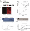
LDH-A is required for PMVECs to sustain aerobic glycolysis and rapid growth. A: PMVECs were infected with a retrovirus (cat. no. 2641), enabling reverse tetracycline-controlled transactivator protein (rtTA) expression. Cells were selected to homogeneity using blasticidin, reinfected with a lentivirus (cat. no. 2894-2), and selected to homogeneity using puromycin. The resulting double-transfection enabled doxycycline-responsive expression of a LDH-A short hairpin RNA (shRNA). Bsr, blasticidin resistance gene; EGFP, enhanced green fluorescent protein; HIV RRE, human immunodeficiency virus Rev response element; IRES EMV, encephalomyocarditis virus internal ribosome entry site; LTR, retro/lentiviral long terminal repeat; PAC, puromycin resistance gene; PSV40, simian virus 40 promoter; PTet, doxycycline-regulated promoter; wPRE, woodchuck hepatitis virus posttranscriptional regulatory element; mir, 5′–3′ flanking sequence derived from the murine mir (micro-RNA gene)-15. B: doxycycline promoted expression of the red fluorescent protein mCherry in PMVECs infected with both retrovirus cat. no. 2641 and lentivirus cat. no. 2894-2 and selected to homogeneity using blasticidin and then puromycin. mCherry fluorescence reveals the uniform doxycycline-responsive shRNA expression. C: Western analysis demonstrates that daily doxycycline treatment decreases LDH-A protein over a 7-day time course, resulting in decreased PMVEC growth (D), glucose consumption (E), and lactate production (F). Whereas doxycycline treatment decreases LDH-A protein (G) and PMVEC growth (H), both LDH-A protein and PMVEC growth are rescued on doxycycline withdrawal. One-way ANOVA was used to assess significance over the 7- or 10-day time course, 2-way ANOVA was used to compare between groups, and Bonferroni post hoc test was performed as needed. *Significantly different (P < 0.05; n = 3). ^Significantly different (P < 0.05) from baseline at day 1.

References

    1. Al-Mehdi AB. Mechanotransduction of shear-stress at the mitochondria. In: Advances in Biochemistry in Health and Disease: Mitochondria: The Dynamic Organelle, edited by Shaffer SW, Suleimon MS. New York: Springer, 2007.
    1. Alexeyev MF, Fayzulin R, Shokolenko IN, Pastukh V. A retro-lentiviral system for doxycycline-inducible gene expression and gene knockdown in cells with limited proliferative capacity. Mol Biol Rep 37: 1987– 1991, 2010 - PMC - PubMed
    1. Alvarez DF, Huang L, King JA, ElZarrad MK, Yoder MC, Stevens T. Lung microvascular endothelium is enriched with progenitor cells that exhibit vasculogenic capacity. Am J Physiol Lung Cell Mol Physiol 294: L419– L430, 2008 - PubMed
    1. Bauer DE, Harris MH, Plas DR, Lum JJ, Hammerman PS, Rathmell JC, Riley JL, Thompson CB. Cytokine stimulation of aerobic glycolysis in hematopoietic cells exceeds proliferative demand. FASEB J 18: 1303– 1305, 2004 - PMC - PubMed
    1. Brand KA, Hermfisse U. Aerobic glycolysis by proliferating cells: a protective strategy against reactive oxygen species. FASEB J 11: 388– 395, 1997 - PubMed

Publication types

MeSH terms

LinkOut - more resources