. 2010 Jul;22(7):2509-26.
doi: 10.1105/tpc.109.069807. Epub 2010 Jul 30.

NENA, a Lotus japonicus homolog of Sec13, is required for rhizodermal infection by arbuscular mycorrhiza fungi and rhizobia but dispensable for cortical endosymbiotic development

Affiliations

NENA, a Lotus japonicus homolog of Sec13, is required for rhizodermal infection by arbuscular mycorrhiza fungi and rhizobia but dispensable for cortical endosymbiotic development

Martin Groth et al. Plant Cell. 2010 Jul.

Abstract

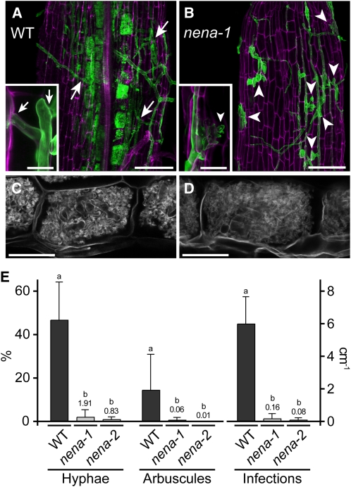
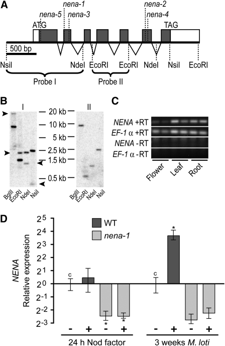
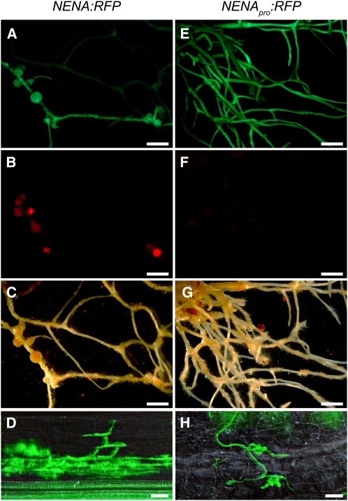
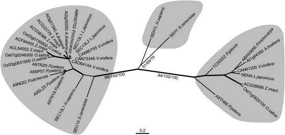
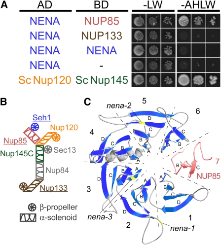
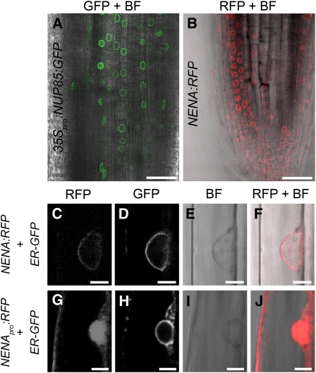
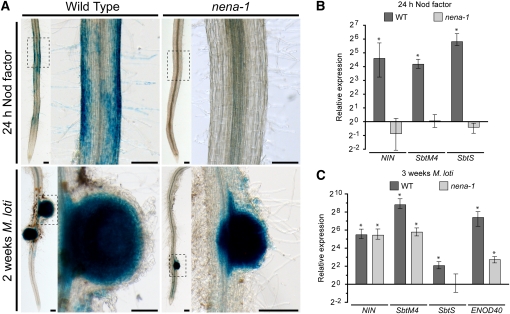
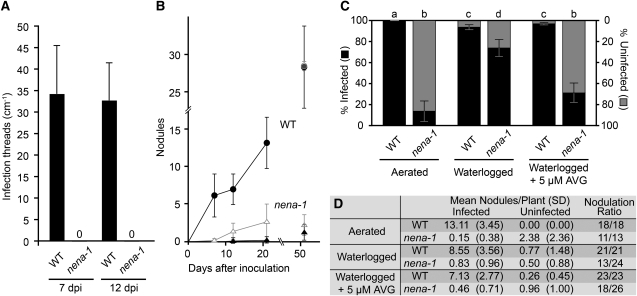
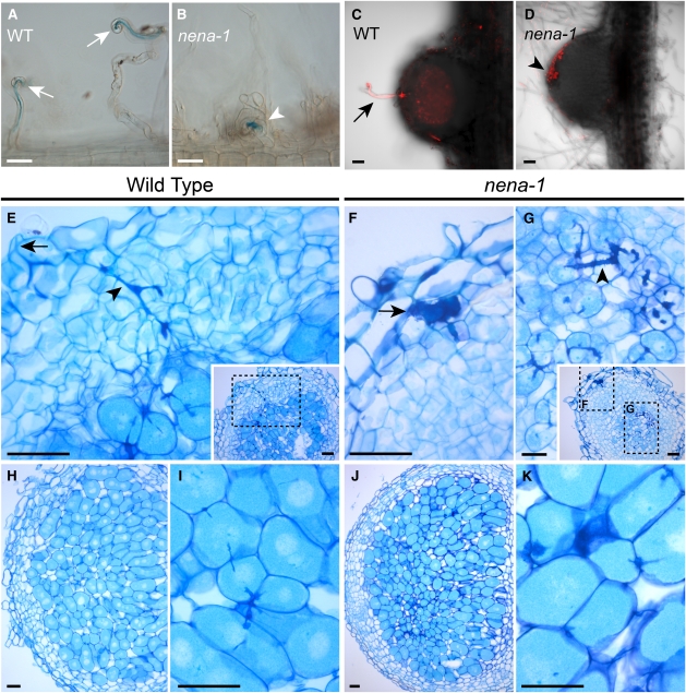
Legumes form symbioses with arbuscular mycorrhiza (AM) fungi and nitrogen fixing root nodule bacteria. Intracellular root infection by either endosymbiont is controlled by the activation of the calcium and calmodulin-dependent kinase (CCaMK), a central regulatory component of the plant's common symbiosis signaling network. We performed a microscopy screen for Lotus japonicus mutants defective in AM development and isolated a mutant, nena, that aborted fungal infection in the rhizodermis. NENA encodes a WD40 repeat protein related to the nucleoporins Sec13 and Seh1. Localization of NENA to the nuclear rim and yeast two-hybrid experiments indicated a role for NENA in a conserved subcomplex of the nuclear pore scaffold. Although nena mutants were able to form pink nodules in symbiosis with Mesorhizobium loti, root hair infection was not observed. Moreover, Nod factor induction of the symbiotic genes NIN, SbtM4, and SbtS, as well as perinuclear calcium spiking, were impaired. Detailed phenotypic analyses of nena mutants revealed a rhizobial infection mode that overcame the lack of rhizodermal responsiveness and carried the hallmarks of crack entry, including a requirement for ethylene. CCaMK-dependent processes were only abolished in the rhizodermis but not in the cortex of nena mutants. These data support the concept of tissue-specific components for the activation of CCaMK.


Figures

Figure 1.
Figure 1.
nena Is Impaired in AM Fungal Infection. (A) to (D) Confocal micrographs of WGA-Alexa Fluor 488-stained AM fungal structures (green in [A] and [B]) associated with wild-type (WT) and nena plants. In the wild type (A), the AM fungus penetrated the outer cell layers (arrows), colonized the root cortex, and formed arbuscules. In nena-1 (B), hyphae grew on the root surface, and balloon-shaped hyphal structures (arrowheads) occurred at aborted infection sites. Insets show infection sites at higher magnification. Root cell walls were stained with propidium iodide and are shown in magenta. Arbuscules formed in nena-2 (D), did not differ from the wild type (C). AM phenotypes of nena-1 and nena-2 did not differ. Images represent observations from more than eight plants per line cocultivated with BEG195 for 3 weeks. (A) and (B) show Z-projections of GFP/RFP overlays; (C) and (D) show the GFP channel. Bars = 100 μm in (A) to (D) and 20 μm in the insets. (E) Mean hyphal colonization (Hyphae, %), arbuscular colonization (Arbuscules, %) per root, and successful infection sites per centimeter per root (Infections) from wild-type and nena plants (n ≥ 4) after 3 weeks of cultivation at 24°C. Small values are shown by numbers above bars. Error bars show sd. Different letters above bars indicate significant differences (P ≤ 0.05, t test) between pairwise comparisons.
Figure 2.
Figure 2.
NENA Is a Single Copy Gene That Is Expressed in Various Tissues and Upregulated in Nodulated Roots. (A) Gene structure of NENA. Closed boxes, open boxes, and triangles represent coding exons, untranslated regions, and introns, respectively. Positions of start and stop codons of the NENA ORF, mutations in different nena alleles, and restriction sites of selected endonucleases are indicated. Braces span the restriction fragments used as probes in DNA gel blot analyses, as referred to in (B). (B) DNA gel blot radiographs of L. japonicus genomic DNA digested with BglII, EcoRI, NdeI, or NsiI hybridized with Probe I or II. Arrowheads mark bands that do not correspond to the genomic context of NENA but are due to partial gene duplication. (C) Expression of NENA in leaves, flowers, and roots (two biological replicates) analyzed by RT-PCR. NENA and the reference gene EF-1α were amplified with (+RT) or without (−RT) preceding reverse transcription. (D) Quantitative PCR analysis of NENA expression in wild-type (WT) and nena-1 roots 24 h after NF treatment or 3 weeks after M. loti inoculation (+). Expression is relative to mock (−) treated wild-type controls (c) and normalized to EF-1α levels. Mean values and se were derived from three biological replicates. Asterisks indicate significant (P < 0.05) differences to control levels.
Figure 3.
Figure 3.
Transgenic Complementation of nena-1. (A) to (D) A. rhizogenes–mediated transformation of nena-1 mutants with genomic NENA including 2 kb of 5′ regulatory sequence fused to RFP (NENA:RFP) led to restoration of RNS ([A] to [C]) and AM establishment (D). (E) to (H) A. rhizogenes–mediated transformation of nena-1 with the 2-kb 5′ regulatory sequence fused to RFP (NENApro:RFP) did not restore RNS ([E] to [G]) and AM (H). Epifluorescence microscopy images show GFP expression in transgenic roots ([A] and [E]) and DsRed expression by M. loti in root nodules ([B] and [F]) using a GFP and a RFP filter, respectively. Corresponding white light illumination images are shown in (C) and (G). Root segments containing AM fungal structures (green) stained with WGA-Alexa Fluor 488 were visualized by DIC/epifluorescence microscopy using a GFP filter ([D] and [H]). Bars = 2 mm in (A) to (C) and (E) to (G) and 40 μm in (D) and (H). [See online article for color version of this figure.]
Figure 4.
Figure 4.
Phylogenetic Relationships of Sec13/Seh1-Like Proteins. The phylogenetic tree includes amino acid sequences from yeast, human, and representative species of bryophytes (Physcomitrella patens), gymnosperms (Picea spp), monocots (Oryza sativa and Z. mays), and dicots (Arabidopsis, L. japonicus, Populus trichocarpa, and Vitis vinifera). Three statistically supported clusters are highlighted: Sec13-related sequences on the left, yeast/human Seh1 in the middle, and plant Seh1-related sequences, including NENA, on the right. Distances correspond to the best-fitting maximum likelihood tree. Branch labels indicate bootstrap values (n = 1000) of maximum likelihood/neighbor-joining/maximum parsimony consensus trees. Thick, medium, and thin branches indicate bootstrap values ≥90, ≥80, and <80 in at least two consensus trees, respectively. Labels at the branch tips include GenBank accession numbers or gene names (accession numbers in Methods). Isoforms from splice variants were excluded from the analysis.
Figure 5.
Figure 5.
NENA Interacts with NUP85 from L. japonicus and Adopts a β-Propeller Structure According to Homology Modeling. (A) Gal4-based yeast two-hybrid assay for interaction between NENA as prey (AD) and NUP85, NUP133, or NENA as bait (BD). The empty bait vector (−) was used as negative control and AD:Sc NUP120 and BD:Sc NUP145 as positive control. Cotransformed yeast was grown in three dilutions on synthetic dropout medium lacking Leu and Trp (-LW) or adenine, His, Leu, and Trp (-AHLW). (B) Schematic representation of the yeast Nup84 subcomplex and the arrangement of its components (Lutzmann et al., 2002). Putative homologs known to be required for root symbioses in L. japonicus are underlined. Color scheme refers to (A) and (C). (C) Ribbon representation of a conceptual β-propeller formed by NENA (blue β-strands, model comprises residues 11 to 317) and the N terminus of NUP85 (red, residues 36 to 94). The model is based on crystal structures of Sc Seh1•Sc Nup85. Individual blades are delimited by dashed lines and numbered. Letters correspond to successive β-strands in each blade. Lack of β-strands in blades 6 and 7 is due to missing template data and incomplete sequence alignment. Positions of mutations in alleles nena-1, -2, and -3 are indicated. [See online article for color version of this figure.]
Figure 6.
Figure 6.
Perinuclear in Vivo Localization of NUP85 and NENA Fusion Proteins. (A) Overlay of fluorescence and bright-field confocal micrographs showing perinuclear green fluorescence in root tip cells expressing 35Spro:NUP85:GFP. (B) Overlay of fluorescence and bright-field confocal micrographs showing perinuclear red fluorescence in root tip cells expressing NENA:RFP and the ER-GFP marker (data not shown). (C) to (F) Confocal micrographs of a rhizodermal cell expressing NENA:RFP ([C] and [F]) and the ER-GFP marker (D). (G) to (J) Confocal micrographs of a rhizodermal cell expressing cytonucleoplasmic NENApro:RFP ([G] and [J]) and the ER-GFP marker (H). Images are from wild-type (A) and nena-1 ([B] to [J]) A. rhizogenes–transformed roots and were acquired in sequential mode at excitationλ = 561 nm/detectionλ = 570 to 630 nm (RFP), excitationλ = 488 nm/detectionλ = 495 to 555nm (GFP) or bright-field (BF). Bars = 40 μm in (A) and (B) and 5 μm in (C) to (J).
Figure 7.
Figure 7.
Rhizodermal Nod Factor Response Is Impaired, Whereas Induction of Symbiosis Genes at Nodule Primordia Is Not Affected, in nena. (A) Bright-field images of X-Gluc incubated roots transformed with NINpro:GUS after NF treatment or inoculation with M. loti. No blue rhizodermal staining was observed in nena-1 roots after NF treatment. Images correspond to Table 3. Boxed regions are shown at higher magnification. Bars = 0.2 mm. (B) and (C) Quantitative PCR analysis of symbiosis gene expression in wild-type and nena-1 roots 24 h after NF treatment (B) or 3 weeks after M. loti inoculation (C). Expression is relative to mock-treated samples and normalized to EF-1α levels. Mean and se were derived from three biological replicates. Asterisks indicate significant (P < 0.05) differences in gene expression between NF or M. loti and mock treatments.
Figure 8.
Figure 8.
Rhizobial Infection of nena Does Not Occur via Root Hairs and Is Promoted by Ethylene. (A) Quantification of root hair ITs 7 and 12 DAI with M. loti expressing DsRed and growth under aerated conditions; no ITs were observed in nena-1. Mean and sd were calculated from ≥19 (nena-1) and ≥14 wild-type (WT) root systems per time point. (B) Nodulation time course during aerated growth conditions after inoculation with M. loti expressing DsRed. Mean and sd were calculated from 13 to 21 nena-1 (triangles) and 12 to 18 wild-type (squares) root systems per time point. Open/gray and closed/black symbols represent total and infected nodules, respectively. If all nodules were infected, only infected nodules are indicated. If all nodules were uninfected, only total nodules are indicated. (C) and (D) Quantification of nodules from wild-type and nena-1 plants cultivated under different conditions 21 DAI with M. loti expressing DsRed. (C) Bars indicate mean percentages of uninfected (gray) and infected (black) nodules per nodulated individual. Error bars indicate se. Different letters above bars indicate significant differences (P ≤ 0.05, t test) between pairwise comparisons. (D) Mean per plant, sd, and number of nodulated plants versus total number of plants per line and treatment (nodulation ratio) are indicated.
Figure 9.
Figure 9.
Rhizobial Microcolonies at the Root Surface of nena-1 Lead to Nodule Formation and Intercellular Entry. (A) and (B) Bright-field DIC images from roots hairs 7 DAI with lacZ-expressing M. loti and 18°C growth temperature. Wild-type (WT) plants show root hair curling (arrows) and ITs, whereas nena-1 mutants display abnormal root hair deformation and occasional colony formation by rhizobia (arrowhead). Images represent observations from more than eight plants per line. Bars = 50 μm. (C) and (D) Confocal z-projections of longitudinal 80-μm tissue sections of a young infected wild-type (C) and an uninfected nena-1 (D) nodule. Images represent samples from 16 DAI/aerated (C) and 21 DAI/waterlogged + 5 μM AVG (D) treatments, corresponding to Figure 8D. (C) DsRed expressing rhizobia (red) have colonized the nodule via an intracellular root hair IT (arrow). (D) An uninfected nodule developed coinciding with accumulation of rhizobia at the root surface (arrowhead). (E) to (K) Thin sections of nodule tissue stained with toluidine blue. (E) Young wild-type nodule with intracellular IT (arrowhead) spanning from the infection site (arrow) into the cortex. (F) and (G) Young nena-1 nodule with a subepidermal infection pocket (arrow) and cortical ITs (arrowhead). Insets in (E) and (G) show respective sections at lower magnification; dashed boxes indicate magnified areas. Longitudinal sections of mature nodules from the wild type (H) or nena-1 (J) and corresponding magnifications ([I] and [K]) showing colonized host cells. Plants were grown under waterlogging conditions and sampled 3 WAI with M. loti R7A. Bars = 50 μm, except (G), where bar = 20 μm.

References

    1. Alber F., Dokudovskaya S., Veenhoff L.M., Zhang W., Kipper J., Devos D., Suprapto A., Karni-Schmidt O., Williams R., Chait B.T., Sali A., Rout M.P. (2007). The molecular architecture of the nuclear pore complex. Nature 450: 695–701 - PubMed
    1. Andriankaja A., Boisson-Dernier A., Frances L., Sauviac L., Jauneau A., Barker D.G., de Carvalho-Niebel F. (2007). AP2-ERF transcription factors mediate Nod factor-dependent Mt ENOD11 activation in root hairs via a novel cis-regulatory motif. Plant Cell 19: 2866–2885 - PMC - PubMed
    1. Ané J.M., et al. (2004). Medicago truncatula DMI1 required for bacterial and fungal symbioses in legumes. Science 303: 1364–1367 - PubMed
    1. Arnold K., Bordoli L., Kopp J., Schwede T. (2006). The SWISS-MODEL workspace: A web-based environment for protein structure homology modelling. Bioinformatics 22: 195–201 - PubMed
    1. Arrighi J.F., et al. (2006). The Medicago truncatula lysine motif-receptor-like kinase gene family includes NFP and new nodule-expressed genes. Plant Physiol. 142: 265–279 - PMC - PubMed

Publication types