. 2010 Aug 17;107(33):14662-7.
doi: 10.1073/pnas.0909432107. Epub 2010 Jul 30.

Cardiac conduction is required to preserve cardiac chamber morphology

Affiliations

Cardiac conduction is required to preserve cardiac chamber morphology

Neil C Chi et al. Proc Natl Acad Sci U S A. .

Abstract

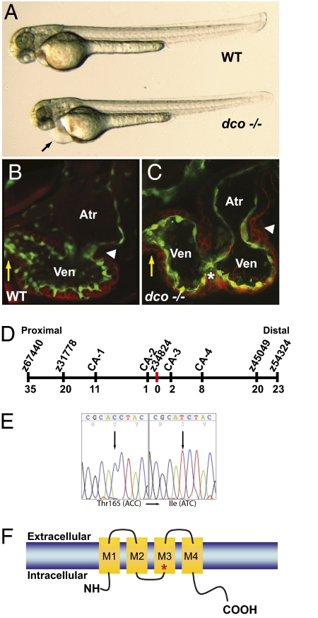
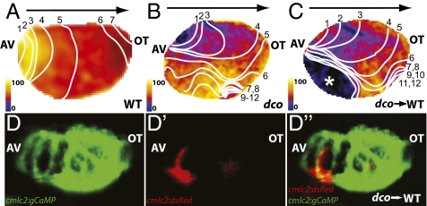
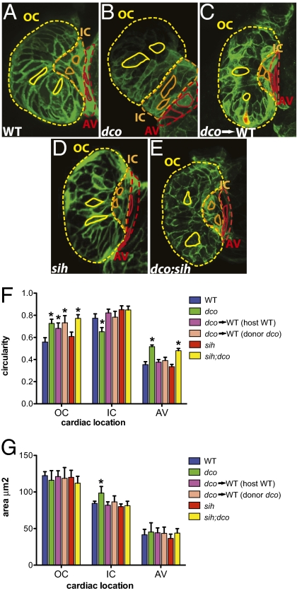
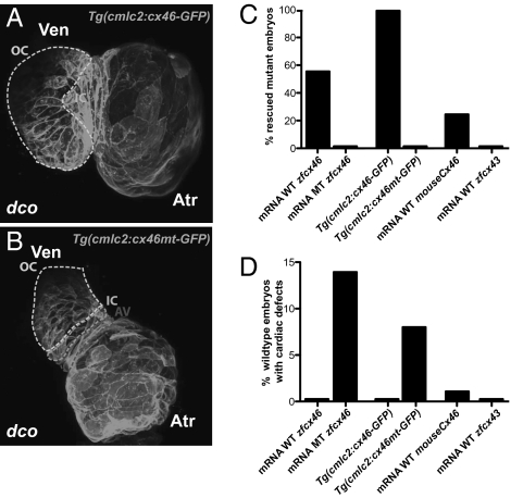
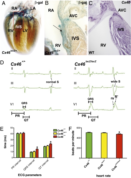
Electrical cardiac forces have been previously hypothesized to play a significant role in cardiac morphogenesis and remodeling. In response to electrical forces, cultured cardiomyocytes rearrange their cytoskeletal structure and modify their gene expression profile. To translate such in vitro data to the intact heart, we used a collection of zebrafish cardiac mutants and transgenics to investigate whether cardiac conduction could influence in vivo cardiac morphogenesis independent of contractile forces. We show that the cardiac mutant dco(s226) develops heart failure and interrupted cardiac morphogenesis following uncoordinated ventricular contraction. Using in vivo optical mapping/calcium imaging, we determined that the dco cardiac phenotype was primarily due to aberrant ventricular conduction. Because cardiac contraction and intracardiac hemodynamic forces can also influence cardiac development, we further analyzed the dco phenotype in noncontractile hearts and observed that disorganized ventricular conduction could affect cardiomyocyte morphology and subsequent heart morphogenesis in the absence of contraction or flow. By positional cloning, we found that dco encodes Gja3/Cx46, a gap junction protein not previously implicated in heart formation or function. Detailed analysis of the mouse Cx46 mutant revealed the presence of cardiac conduction defects frequently associated with human heart failure. Overall, these in vivo studies indicate that cardiac electrical forces are required to preserve cardiac chamber morphology and may act as a key epigenetic factor in cardiac remodeling.


Conflict of interest statement

The authors declare no conflict of interest.

Figures

Fig. 1.
Fig. 1.
dococs226 Mutants exhibit pericardial edema and cardiac failure due to dysynchronous contraction of the ventricle. (A) Brightfield micrographs of 48 hpf WT (WT) and dcos226 mutant (dco −/−) embryos. Black arrow points to pericardial edema. (B and C) Confocal micrographs of 60 hpf Tg(flk1:EGFP)s843 (B) WT and (C) dcos226 mutant hearts (endothelium green) stained with rhodamine phalloidin (myocardium red). dco Mutant ventricles fail to contract coordinately and exhibit disorganized chamber walls. White arrowheads point to atrioventricular canal; yellow arrows point to outflow tract; white asterisk denotes aberrant ventricular wall contraction. (D) Genetic map of the dco region. Numbers below SSLP markers indicate recombination events out of 922 diploid embryos examined. (E) Sequencing of cx46 cDNA revealed a C-to-T change at base pair 494 in the s226 mutant allele, resulting in a Thr-to-Ile substitution at residue 165. (F) Schematic diagram illustrating modular structure of Cx46. A connexon consists of four transmembrane domains (M1–M4). The M3 domain is the major pore-lining domain. Red asterisk marks site affected by s226 mutation.
Fig. 2.
Fig. 2.
Optical mapping/calcium imaging of dococs226 mutants reveals disrupted cardiac conduction. (A–C) Optical maps of calcium excitation during a single cardiac cycle, represented by isochronal lines every 20 ms in 60 hpf embryos carrying the Tg(cmlc2:gCaMP)s878 reporter and (A) WT at the dco locus (WT), (B) dcos226 homozygous mutant (dco), or (C) WT with dcos226 mutant cardiomyocytes carrying the Tg(cmlc2:dsRed)s879 reporter in the ventricle (dco → WT). Black arrow shows normal direction of ventricular cardiac conduction. Numbers indicate temporal sequence of calcium excitation. Color bar chart fluorescence intensity changes on a scale of 0 to 100. Cardiac conduction across the WT ventricle proceeds uniformly from the atrioventricular canal (AV) to the outflow tract (OT), whereas cardiac conduction travels aberrantly through the dcos226 mutant ventricle. Transplanted dcos226 mutant cardiomyocytes disrupt the organized AV to OT conduction in WT ventricle. White asterisk marks location of transplanted dco mutant cardiomyocytes in WT ventricle, as illustrated in D′ and D′′. (D and D′′) Fluorescent micrographs of the dco → WT mosaic heart imaged in C. (D) Green fluorescence shows the WT host ventricle; (D′) donor-derived dcos226 mutant cardiomyocytes expressing Tg(cmlc2:dsRed)s879. (D′′) Overlay of green and red fluorescence images reveals that the donor-derived dcos226 mutant cardiomyocytes are located at the outer curvature of the ventricle near the AV canal, where cardiac conduction is disrupted in the WT host ventricle (C).
Fig. 3.
Fig. 3.
Cardiac conduction, independent of hemodynamic flow or cardiac contraction, is required for cardiomyocyte morphogenesis. (A–E) Confocal images of 60 hpf embryos carrying the Tg(cmlc2:ras-eGFP)s883 reporter and (A) WT at the dco locus (WT), (B) dcos226 homozygous mutant (dco), (C) WT with dcos226 mutant cardiomyocytes carrying the Tg(cmlc2:dsRed)s879 reporter in the ventricle (dco → WT), (D) sih homozygous mutant (sih), or (E) dco;sih double homozygous mutant (dco;sih). Outer curvature (OC), inner curvature (IC), and atrioventricular canal (AV) are outlined in yellow, orange, and red dashed lines, respectively. Representative cardiomyocyte shapes for each area are outlined with solid lines. Bar graphs represent (F) cell morphology/circularity and (G) surface area measurements of Tg(cmlc2:ras-eGFP)s883 cardiomyocytes at 60 hpf from the outer and inner curvatures of the ventricle as well as the AV canal. Bar height indicates mean for a dataset; error bars indicate SE. *Statistically significant differences compared with WT (P < 0.001). Defects in electrical conduction result in aberrant cardiomyocyte morphogenesis.
Fig. 4.
Fig. 4.
dco Cardiac phenotype is rescued by RNA injection of Cx46 orthologs as well as by myocardial specific expression of zebrafish cx46. (A and B) Myocardial specific expression of WT [Tg(cmlc2:cx46-GFPs882)] and mutant [Tg(cmlc2:cx46mt-GFPs925)] zebrafish Cx46-GFP fusion proteins lead to cell membrane enrichment. Outer curvature (OC), inner curvature (IC), and atrioventricular canal (AV) are outlined by dashed lines. Atr, atrium; Ven, ventricle. (C) Injection of WT zebrafish (WT zfcx46) or mouse Cx46 (mouseCx46) mRNA as well as myocardial specific expression of WT zebrafish Cx46-GFP fusion proteins [Tg(cmlc2:cx46-GFPs882)] rescued the dco cardiac phenotype. On the other hand, injection of zebrafish cx46s226 (MT zfcx46) or cx43 (zfcx43) mRNA as well as myocardial specific expression of zebrafish Cx46s226-GFP [Tg(cmlc2:cx46mt-GFPs925)] failed to rescue the dco cardiac phenotype. (D) However, injection of zebrafish cx46s226 mRNA or myocardial specific expression of zebrafish Cx46s226-GFP [Tg(cmlc2:cx46mt-GFPs925)] in WT embryos resulted in dco cardiac phenotypes. In addition, RNA overexpression of WT murine Cx46 caused a dco cardiac phenotype in a small percentage of WT embryos.
Fig. 5.
Fig. 5.
Murine Cx46 is expressed in the fast cardiac conduction system, and its disruption results in cardiac conduction defects. (A) Whole-mount β-gal staining of Cx46lacZ/+ postnatal mice (P3). Cx46lacZ/+ cells are detected in the atrium, ventricle, atrioventricular canal (AVC), and interventricular septum (IVS). Red box outlines the IVS and AVC regions. (B) Section analysis of region outlined by red box in A containing IVS and AVC reveals Cx46lacZ/+ cells in subendocardium of IVS where His-Purkinje fibers reside (black arrow). (C) Section RNA in situ hybridization of endogenous Cx46 expression in WT CD1 P3 mice is consistent with X-gal staining of Cx46lacZ/+ cardiac tissue. (D) Representative three-lead electrocardiogram (ECG) recordings from WT and Cx46lacZ/lacZ (null) mice. Cx46 null ECGs exhibited prolonged QRS and QT intervals, suggesting an intraventricular conduction delay. Cx46 null ECGs also exhibited an rSR′ morphology in lead V1 and a wide S wave in leads II and III, indicating uncoordinated ventricular activation. Bar graphs represent (E) ECG measurements (PR, QRS intervals, and QT), and (F) heart rate of Cx46+/+, Cx46lacZ/+, and Cx46lacZ/lacZ hearts. Bar height indicates mean for datasets; error bars indicate SE (Table S2). *Statistically significant differences between phenotypically WT (Cx46+/+ and Cx46lacZ/+) and mutant (Cx46lacZ/lacZ) hearts (P < 0.05). LA, left atrium; LV, left ventricle; RA, right atrium; RV, right ventricle.

References

    1. Olson EN. Gene regulatory networks in the evolution and development of the heart. Science. 2006;313:1922–1927. - PMC - PubMed
    1. Auman HJ, et al. Functional modulation of cardiac form through regionally confined cell shape changes. PLoS Biol. 2007;5:e53. - PMC - PubMed
    1. Bartman T, et al. Early myocardial function affects endocardial cushion development in zebrafish. PLoS Biol. 2004;2:E129. - PMC - PubMed
    1. Hove JR, et al. Intracardiac fluid forces are an essential epigenetic factor for embryonic cardiogenesis. Nature. 2003;421:172–177. - PubMed
    1. Radisic M, et al. Functional assembly of engineered myocardium by electrical stimulation of cardiac myocytes cultured on scaffolds. Proc Natl Acad Sci USA. 2004;101:18129–18134. - PMC - PubMed

Publication types

MeSH terms