. 2010 Nov;38(21):7526-37.
doi: 10.1093/nar/gkq664. Epub 2010 Jul 31.

Dynamics of a novel centromeric histone variant CenH3 reveals the evolutionary ancestral timing of centromere biogenesis

Affiliations

Dynamics of a novel centromeric histone variant CenH3 reveals the evolutionary ancestral timing of centromere biogenesis

Manu Dubin et al. Nucleic Acids Res. 2010 Nov.

Abstract

The centromeric histone H3 variant (CenH3) serves to target the kinetochore to the centromeres and thus ensures correct chromosome segregation during mitosis and meiosis. The Dictyostelium H3-like variant H3v1 was identified as the CenH3 ortholog. Dictyostelium CenH3 has an extended N-terminal domain with no similarity to any other known proteins and a histone fold domain at its C-terminus. Within the histone fold, α-helix 2 (α2) and an extended loop 1 (L1) have been shown to be required for targeting CenH3 to centromeres. Compared to other known and putative CenH3 histones, Dictyostelium CenH3 has a shorter L1, suggesting that the extension is not an obligatory feature. Through ChIP analysis and fluorescence microscopy of live and fixed cells, we provide here the first survey of centromere structure in amoebozoa. The six telocentric centromeres were found to mostly consist of all the DIRS-1 elements and to associate with H3K9me3. During interphase, the centromeres remain attached to the centrosome forming a single CenH3-containing cluster. Loading of Dictyostelium CenH3 onto centromeres occurs at the G2/prophase transition, in contrast to the anaphase/telophase loading of CenH3 observed in metazoans. This suggests that loading during G2/prophase is the ancestral eukaryotic mechanism and that anaphase/telophase loading of CenH3 has evolved more recently after the amoebozoa diverged from the animal linage.


Figures

Figure 1.
Figure 1.
The Dictyostelium centromeric histone H3 variant. (A) Dictyostelium H3a and DdCenH3 (H3v1) are highlighted in orange on the left. Below the alignment is a schematic presentation of the domain organization of histone H3, with the N-terminal α-helix (αN), α-helixes 1–3 (α1–3) and loops 1 and 2 (L1, L2) indicated. L1 and α2, which have been shown to be responsible for targeting of CenH3 to centromeres are shaded green. Hs, Homo sapiens; At, Arabidopsis thaliana; Dd, Dictyostelium discodeum; Eh, Entamoeba histolytica; Sc, Saccharomyces cerevisiae; Xl, Xenopus laevis; Ce, Caenorhabditis elegans; Dt, Drosophila teissieri; Gi, Giardia intestinalis. (B) The organization of the centromeres was examined in fixed cells expressing GFP-DdCenH3 and immunostained with the centrosome marker DdCP224 at the indicated stages of the cell cycle. (C) Prophase of a diploid cell immunostained with DdCP224 and expressing GFP-DdCenH3 displays up to 12 GFP-DdCenH3 labelled foci presumably corresponding to the 12 centromeres. Scale bar = 1 µm.
Figure 2.
Figure 2.
Centromere behaviour during mitosis. (A) Cells expressing GFP-DdCenH3 and labelled with the heterochromatin marker H3K9me3. DNA is stained with DAPI. (B) Cross sections through cells at different stages of the mitotic cycle show that GFP-DdCenH3 (green) is slightly closer to the leading edge of the separating chromatids than the heterochromatin marker H3K9me3 (red). Scale bar = 1 µm.
Figure 3.
Figure 3.
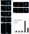
Dictyostelium centromeres contain DIRS-1. (A) FISH on cells at different stages of the cell cycle using a directly FITC-labelled probe for DIRS-1. (B) Combined Immuno-FISH showing colocalization HA-tagged DdCenH3 and DIRS1 retrotransposon. Scale bar = 1 µm. (C) ChIP of GFP-DdCenH3. Real-time qPCR was performed with primers against the actively transcribed actin, coronin and rasG genes or the DIRS-1 and skipper retrotransposons. noAb = no antibody control.
Figure 4.
Figure 4.
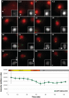
Loading of GFP-DdCenH3 onto centromeres. (A) Cells co-expressing GFP-DdCenH3 and the cell cycle marker RFP-PCNA were fixed and the normalized intensity of the GFP-DdCenH3 signal at the centromeres at different stages of the cell cycle was plotted. (B) Time-lapse images of a cell with three nuclei co-expressing GFP-DdCenH3 and RFP-H2AX. Images were acquired every 20 s and the overlay of the GFP and RFP signals from every third acquisition (every 60 s) is displayed. The GFP signal for one centromere at each time point (indicated with an arrow) is shown as an insert in grey scale. Signal intensities above a certain arbitrary threshold are shaded red. The average signal intensities from the three nuclei averaged over three time points (1 min) are plotted together with the standard error. The bar above the charts indicates the stage of the cell cycle Scale bar = 1 µm. Note that the values in the graph are calculated from a minimum of nine individual data points and do not strictly correspond to the single-intensity data displayed in the inserts.
Figure 5.
Figure 5.
S-phase Behaviour of GFP-DdCenH3. Time-lapse images of cells co-expressing GFP-DdCenH3 and RFP-PCNA starting from telophase. Images were acquired every 90 s and the overlay of the GFP an RFP signals is displayed. The time elapsed since the start of the acquisition is indicated in the upper left hand corner of each frame. The RFP signal for one centromere at each time point (indicated with an asterisk) is shown as an insert in greyscale. The strongly stained spot of RFP-PCNA characteristic of late S-phase is indicated with an arrow. The intensity of the GFP-DdCenH3 signal is averaged over three time points (4.5 min) and plotted below. The bar above the chart indicates the stage of the cell cycle. Scale bar = 1 µm.

References

    1. Gieni RS, Chan GK, Hendzel MJ. Epigenetics regulate centromere formation and kinetochore function. J. Cellular Biochem. 2008;104:2027–2039. - PubMed
    1. Tyler-Smith C, Floridia G. Many paths to the top of the mountain, diverse evolutionary solutions to centromere structure. Cell. 2000;102:5–8. - PubMed
    1. Ekwall K. Epigenetic control of centromere behavior. Annu. Rev. Genet. 2007;41:63–81. - PubMed
    1. Cooper JL, Henikoff S. Adaptive evolution of the histone fold domain in centromeric histones. Mol. Biol. Evol. 2004;21:1712–1718. - PubMed
    1. Malik HS, Henikoff S. Adaptive evolution of cid, a centromere-specific histone in Drosophila. Genetics. 2001;157:1293–1298. - PMC - PubMed

Publication types