. 2010 Nov;38(21):7749-63.
doi: 10.1093/nar/gkq660. Epub 2010 Jul 31.

HEXIM1 targets a repeated GAUC motif in the riboregulator of transcription 7SK and promotes base pair rearrangements

Affiliations

HEXIM1 targets a repeated GAUC motif in the riboregulator of transcription 7SK and promotes base pair rearrangements

Isabelle Lebars et al. Nucleic Acids Res. 2010 Nov.

Abstract

7SK snRNA, an abundant RNA discovered in human nucleus, regulates transcription by RNA polymerase II (RNAPII). It sequesters and inhibits the transcription elongation factor P-TEFb which, by phosphorylation of RNAPII, switches transcription from initiation to processive elongation and relieves pauses of transcription. This regulation process depends on the association between 7SK and a HEXIM protein, neither isolated partner being able to inhibit P-TEFb alone. In this work, we used a combined NMR and biochemical approach to determine 7SK and HEXIM1 elements that define their binding properties. Our results demonstrate that a repeated GAUC motif located in the upper part of a hairpin on the 5'-end of 7SK is essential for specific HEXIM1 recognition. Binding of a peptide comprising the HEXIM Arginine Rich Motif (ARM) induces an opening of the GAUC motif and stabilization of an internal loop. A conserved proline-serine sequence in the middle of the ARM is shown to be essential for the binding specificity and the conformational change of the RNA. This work provides evidences for a recognition mechanism involving a first event of induced fit, suggesting that 7SK plasticity is involved in the transcription regulation.


Figures

Figure 1.
Figure 1.
7SK, HP1 and HEXIM1. (A) Schematic representation of the inhibitory 7SKsnRNP complex. (B) Domain organization of HEXIM1 protein. The central NLS overlaps with the 7SK RNA-binding domain (ARM). (C) SHAPE analysis of the 5′ region of 7SK in full length context (7SK lanes) or isolated (HP1 lanes). Primer extension analysis of 1M7 reactions (+) in three different buffers (A: 50 mM Na HEPES pH 7.6, 6 mM MgCl2, 0.25 mM EDTA; B: 50 mM Na HEPES pH 7.6, 6 mM MgCl2, 0.25 mM EDTA, 200 mM KCl; C: 100 mM Na HEPES pH 8.0, 6 mM MgCl2, 100 mM NaCl) or controls without reactant (−) were migrated in parallel with sequencing reactions (lanes G, C, A, U, identification on the left) to map the single-stranded, 1M7-modified nucleotides, which are identified on the right.
Figure 2.
Figure 2.
Determination of the secondary structure of the HP1 (24–87) hairpin. (A) Sequence of HP1 used for NMR studies. (B) Imino/imino protons region of a NOESY spectrum recorded at 10°C in 90/10 H2O/D2O with a mixing time of 300 ms. Dashed lines represent NH/NH sequential assignment (top). Regions of SOFAST-HMQC, recorded at natural abundance at 10°C, corresponding to the 15N guanosine imino protons (middle) and to 15N uridine imino groups (bottom).
Figure 3.
Figure 3.
Mapping of the interaction between HP1 and RNA-binding domain of HEXIM1 revealed by NMR. (A) Titration of HP1 RNA by ARM peptide. Spectra for free RNA (top) and for HP1:ARM complexes at ratios of 1:0.7 and 1:1.3 are shown. The assignment of imino proton is based on analysis of NOESY experiments. Assignments of G73 and G74 resonances is indicated by italic characters as they can be inverted. Broken lines indicate imino proton that undergo chemical shift changes. (B) Imino/imino protons regions of NOESY spectra recorded at 10°C in 90/10 H2O/D2O with a mixing time of 300 ms. Correlations U84G26, U44G64 and U66G42 are observable in the free HP1 (top). U44G64 disappears in the HP1:ARM complex while U66G42 is shifted (middle). The same effect is observed in the complex HP1:ARM-NLS (bottom). (C) Summary of the effects observed on HP1 upon interaction. The opening of base pairs is indicated with stars. The stabilization of base pairs is indicated with squares. Residues for which imino protons undergo chemical shifts changes upon addition of peptide are highlighted with circles.
Figure 4.
Figure 4.
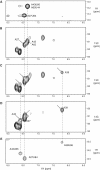
Effect of binding on non-exchangeable protons. (A) 1H NOESY spectrum showing imino/H2 correlations of the free HP1 at 20°C, (B) 1H-13C HSQC spectrum showing the aromatic H2–C2 correlations of the free HP1 at 20°C, (C) 1H-13C HSQC spectrum showing the aromatic H2–C2 correlations of HP1 at 20°C at a ratio 0.5:1 peptide/RNA, (D) 1H-13C HSQC spectrum showing the aromatic H2–C2 correlations of HP1 at 20°C with a ratio 1.3:1 peptide/RNA and (E) 1H NOESY spectrum showing imino/H2 correlations of HP1 at 20°C with a ratio 1.3/1 peptide/RNA. Correlation peaks that appear upon titration are indicated by arrows.
Figure 5.
Figure 5.
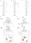
Effect of mutations in the ARM motif on the binding to HP1. (A) Sequences of ARM motif and its mutants. Mutations are indicated with bold letters. Regions corresponding to imino protons involved in AU pairs of HP1 are shown upon titration by S158C (B), P157G (C) and P157K (D).
Figure 6.
Figure 6.
Role of bulges on the interaction with RNA-binding domain of HEXIM1. (A) Secondary structure of HP1ΔU4041 in which U40 and U41 have been deleted. (B) Secondary structure of HP1ΔU63 in which U63 had been deleted. (C) Secondary structure of HP1dm in which mutation GAUC/GAUC to GGCC/GGCC was introduced. Mutations are highlighted in red. (D) Titration of HP1ΔU4041 RNA by ARM domain. The assignment of imino proton based on the analysis of NOESY experiment is indicated for the free RNA (top) and the RNA:ARM complex (middle). Broken lines indicate imino proton that undergo chemical shift changes. Imino/imino protons region of a NOESY spectrum recorded at 10°C in 90/10 H2O/D2O with a mixing time of 300 ms is represented (bottom). Correlations U84G26, U44G64 and U66G42 are observable in the free HP1ΔU4041 (black) and remain unchanged in the bound RNA (red) (E) Titration of HP1ΔU63 RNA by the peptide. The assignment of imino proton based on the analysis of NOESY experiment is indicated for the free RNA (top) and the complex (middle). Broken lines indicate imino proton that undergo chemical shift changes. Imino/imino protons region of a NOESY spectrum recorded at 10°C in 90/10 H2O/D2O with a mixing time of 300 ms is represented (bottom). Correlations U84G26, U44G64 and U66G42 are observable in the free HP1ΔU63 (black) and remain unchanged in the bound RNA (red).
Figure 7.
Figure 7.
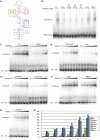
Functional impact of the 7SK mutations on the full-length HEXIM binding. (A) Mutations introduced into the 24–87 region of 7SK. (B) EMSA of several mutated full-length radioactively labeled 7SK at one concentration (0.4 µM) of full-length HEXIM1. (C–G) EMSA of mutated HP1-long (1–108) variants at increasing concentrations (0.1–1.2 µM) of full-length HEXIM1. The concentration of HEXIM1 was maintained low to avoid non-specific binding, a known tendency of HEXIM (70), that leads to the appearance of multiple bands. (H) Recapitulation of full length HEXIM1 binding of all HP1-long mutants. To simplify the figure some mutations were not represented. These are: HP1AU4344 and HP1AU6566 (which behave like HP1dm and ΔU63, respectively), and U28C, G83A, U32C and G79A (which behave like WT).

References

    1. Zieve G, Benecke BJ, Penman S. Synthesis of two classes of small RNA species in vivo and in vitro. Biochemistry. 1977;16:4520–4525. - PubMed
    1. Gurney T, Jr, Eliceiri GL. Intracellular distribution of low molecular weight RNA species in HeLa cells. J. Cell Biol. 1980;87:398–403. - PMC - PubMed
    1. Murphy S, Di Liegro C, Melli M. The in vitro transcription of the 7SK RNA gene by RNA polymerase III is dependent only on the presence of an upstream promoter. Cell. 1987;51:81–87. - PubMed
    1. Kruger W, Benecke BJ. Structural and functional analysis of a human 7 S K RNA gene. J. Mol. Biol. 1987;195:31–41. - PubMed
    1. Wassarman DA, Steitz JA. Structural analyses of the 7SK ribonucleoprotein (RNP), the most abundant human small RNP of unknown function. Mol. Cell Biol. 1991;11:3432–3445. - PMC - PubMed

Publication types