. 2010 Sep 15;29(18):3108-17.
doi: 10.1038/emboj.2010.180. Epub 2010 Jul 30.

The key function of a conserved and modified rRNA residue in the ribosomal response to the nascent peptide

Affiliations

The key function of a conserved and modified rRNA residue in the ribosomal response to the nascent peptide

Nora Vázquez-Laslop et al. EMBO J. .

Abstract

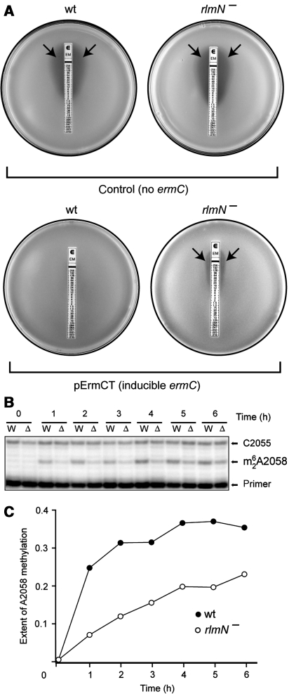
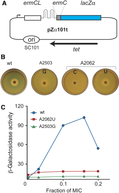
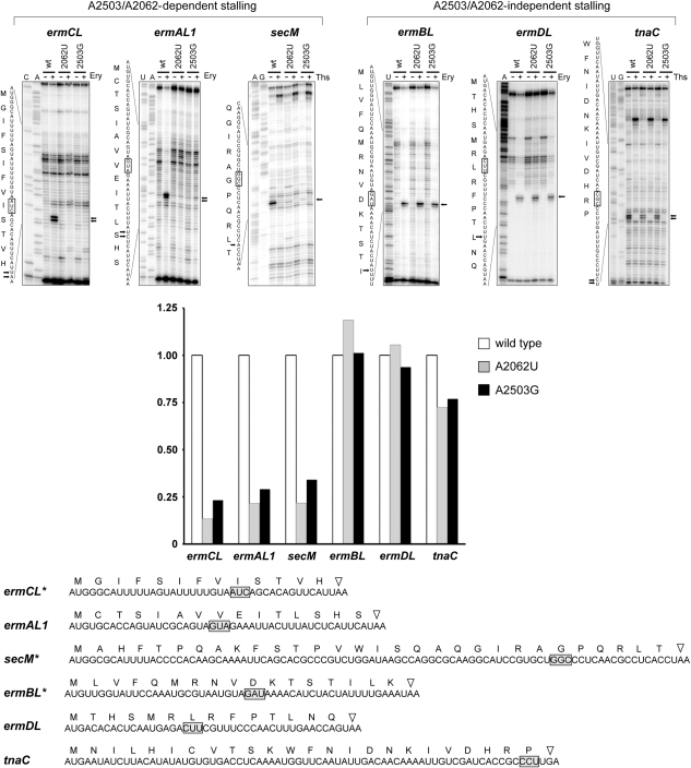
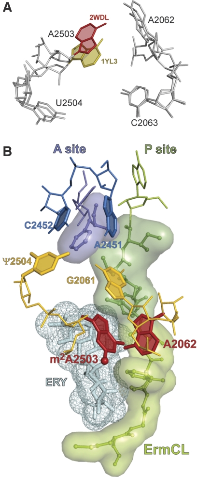
The ribosome is able to monitor the structure of the nascent peptide and can stall in response to specific peptide sequences. Such programmed stalling is used for the regulation of gene expression. The molecular mechanisms of the nascent-peptide recognition and ribosome stalling are unknown. We identified the conserved and posttranscriptionally modified 23S rRNA nucleotide m(2)A2503 located at the entrance of the ribosome exit tunnel as a key component of the ribosomal response mechanism. A2503 mutations abolish nascent-peptide-dependent stalling at the leader cistrons of several inducible antibiotic resistance genes and at the secM regulatory gene. Remarkably, lack of the C2 methylation of A2503 significantly function induction of expression of the ermC gene, indicating that the functional role of posttranscriptional modification is to fine-tune ribosome-nascent peptide interactions. Structural and biochemical evidence suggest that m(2)A2503 may act in concert with the previously identified nascent-peptide sensor, A2062, in the ribosome exit tunnel to relay the stalling signal to the peptidyl transferase centre.


Conflict of interest statement

The authors declare that they have no conflict of interest.

Figures

Figure 1
Figure 1
Nascent peptide in the ribosome exit tunnel. (A) The structure of the inducible ermC operon where the ermC gene is preceded by a regulatory ORF ermCL. Drug- and nascent-peptide-dependent ribosome stalling at ermCL ORF changes the conformation of the mRNA intergenic region (schematically shown as a two-hairpin structure), thereby releasing translational attenuation of ermC. (B, C) Erythromycin and the ErmCL nascent peptide in the ribosome exit tunnel (viewed from the PTC down the tunnel). In the vacant tunnel (B), the nascent-peptide sensor, A2062, is free to rotate into the tunnel lumen. Binding of antibiotic (‘ERY') narrows the tunnel (C). In the constricted tunnel, the ErmCL nascent peptide drives A2062 toward the tunnel wall, where it comes into close proximity to A2503. (D) Conformational flexibility of A2062. The orientations of the A2062 base are shown for the apo structure of the Haloarcula marismortui 50S ribosomal subunit (blue) (PDB accession number 3CC2) (Blaha et al, 2008) and for the 50S subunit complexed with a transition state analog (biege) (1VQ7) (Schmeing et al, 2005). The A2503 base is coloured red. A possible hydrogen bond between A2062 and A2503 is indicated by a dashed line.
Figure 2
Figure 2
The effects of indigenous posttranscriptional modification of A2503 upon ermC induction. (A) The E-test analysis of erythromycin resistance of the E. coli cells that either do not carry the ermC gene (control) or carry a plasmid pErmCT with inducible ermC. The rlmN+ cells (wt) or rlmN cells (lacking posttranscriptional modification of A2503) were plated onto agar plates and overlaid with an E-strip containing a gradient of erythromycin concentration. Inhibition of cell growth is manifested as a clear zone around the strip (arrows). (B) Primer-extension analysis of the induction of 23S rRNA modification by ErmC upon exposure of cells to erythromycin. Exponential wild-type (W) or rlmN (Δ) cells carrying inducible ermC were induced with 32 μg/ml erythromycin, and the extent of ErmC-catalysed A2058 dimethylation in 23S rRNA at specified time points was analysed by primer extension. In the presence of ddGTP terminator, reverse transcriptase stops at the dimethylated A2058 but advances to C2055 when A2058 remains unmodified. (C) Quantification of the intensities of the primer-extension bands on the gel shown in (B), representing the fraction of the modified 23S rRNA.
Figure 3
Figure 3
The effect of the A2503 and A2062 mutations upon induction of the ermC-based reporter. (A) The structure of the ermC-based pZα101t reporter. A portion of the lacZ gene encoding the β-galactosidase α-peptide is fused to the first two codons of ermC. Ribosome stalling at the ermCL ORF induces expression of the ermC-lacZα fusion. (B) Drug-diffusion induction of the pZα101t reporter in E. coli cells expressing wild-type or mutant ribosomes. The plates contain a lawn of SQK15/pZα101t cells grown on the surface of LB agar plates supplemented with ampicillin, tetracycline, IPTG, and X-gal. Expression of the reporter is induced by diffusion of erythromycin from the paper disk containing 0.5 mg of the drug. (C) β-Galactosidase activity (nmol/min/mg) in the wild-type and mutant SQK15 cells containing the pZα101t reporter.
Figure 4
Figure 4
Toeprinting analysis of the effects of rRNA mutations on nascent-peptide-dependent ribosome stalling. The regulatory ORF templates were translated in a cell-free translation system under either the nonstalling or stalling conditions, and formation of the stalled complex was monitored by primer extension. Ribosome stalling signals on the gels and corresponding sequences are indicated by arrows. The codon located in the P site of the stalled ribosome is boxed. Addition of erythromycin (Ery) is necessary to cause ribosome stalling during translation of the leader erm templates (ermCL, ermAL1, ermBL, and ermDL). Thiostrepton (Ths), an inhibitor of translation, was added to the indicated reactions directed by the secM and tnaC templates to demonstrate that appearance of toeprint signals on these cistrons depends on their active translation. Sequencing lanes are marked. Quantification of the normalized relative intensity of the stalling signal band is shown in the bar graph. The complete nucleotide sequences of the ORFs used in cell-free translation and the amino-acid sequences of the encoded peptides are shown below the gels. Stop codons are indicated by triangles. The ORFs marked with asterisks have been modified from the original wild-type versions by truncating the 5′ end (secM) or 3′ end (ermCL and ermBL).
Figure 5
Figure 5
Molecular mechanism of the translation arrest induced by the presence of antibiotic and stalling nascent peptide in the ribosome exit tunnel. (A) Possible syn–anti rotation of the A2503 base. The orientation of A2503 in different crystallographic complexes of T. thermophilus 70S ribosome (PDB accession numbers 2WDL and 1YL3 for the syn and anti conformations, respectively) (Jenner et al, 2005; Voorhees et al, 2009). (B) Molecular interactions leading to programmed ribosome stalling. The 9-amino-acid ErmCL nascent peptide was modelled in the exit tunnel of the T. thermophilus 70S ribosome (pdb accession number 2WDL) according to Tu et al (2005) and energy minimized. Erythromycin was docked by aligning the T. thermophilus structure, with the structure of H. marismortui 50S subunit complexed with erythromycin (1YI2; Tu et al, 2005). In the ribosome stalled at the ermCL ORF, the 9-amino-acid ErmCL nascent peptide (green, sticks and semitransparent surface) esterifies tRNA bound in the ribosomal P site (only the 3′ adenosine of the peptidyl-tRNA is shown). The presence of erythromycin (cyan, sticks and mesh) in the tunnel constrains the peptide placement, compelling the critical C-terminal sequence of the peptide (dark green) to come into direct contact with the A2062 base (red) and forcing it into a conformation where it would clash with m2A2503 (red). Reorientation of m2A2503, possibly facilitated by posttranscriptional methylation at C2 (ball), is then communicated to the PTC active site. Change in the position of G2061 and/or U2504 (pseudouridine, Ψ, in E. coli) (both shown in gold) will likely change the opening of the A2451/C2452 (blue) A-site cavity and prevent proper accommodation of the aminoacyl moiety of the A-site aminoacyl-tRNA (purple).

References

    1. Asai T, Zaporojets D, Squires C, Squires CL (1999) An Escherichia coli strain with all chromosomal rRNA operons inactivated: complete exchange of rRNA genes between bacteria. Proc Natl Acad Sci USA 96: 1971–1976 - PMC - PubMed
    1. Baba T, Ara T, Hasegawa M, Takai Y, Okumura Y, Baba M, Datsenko KA, Tomita M, Wanner BL, Mori H (2006) Construction of Escherichia coli K-12 in-frame, single-gene knockout mutants: the Keio collection. Mol Syst Biol 2: 2006.0008 - PMC - PubMed
    1. Bailey M, Chettiath T, Mankin AS (2008) Induction of ermC expression by ‘non-inducing' antibiotics. Antimicrob Agents Chemother 52: 866–874 - PMC - PubMed
    1. Ban N, Nissen P, Hansen J, Moore PB, Steitz TA (2000) The complete atomic structure of the large ribosomal subunit at 2.4 A resolution. Science 289: 905–920 - PubMed
    1. Bayfield MA, Dahlberg AE, Schulmeister U, Dorner S, Barta A (2001) A conformational change in the ribosomal peptidyl transferase center upon active/inactive transition. Proc Natl Acad Sci USA 98: 10096–10101 - PMC - PubMed

Publication types

MeSH terms

LinkOut - more resources