. 2010 Aug 26;466(7310):1120-4.
doi: 10.1038/nature09296. Epub 2010 Aug 1.

A ribosome-associating factor chaperones tail-anchored membrane proteins

Affiliations

A ribosome-associating factor chaperones tail-anchored membrane proteins

Malaiyalam Mariappan et al. Nature. .

Abstract

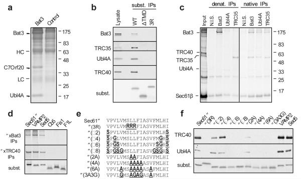
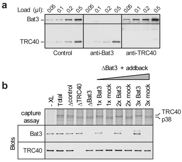
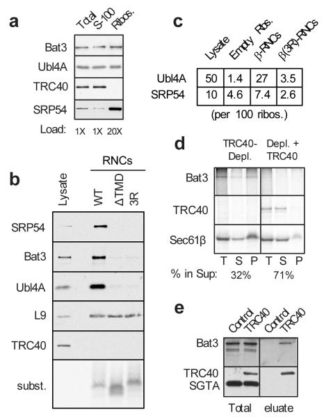
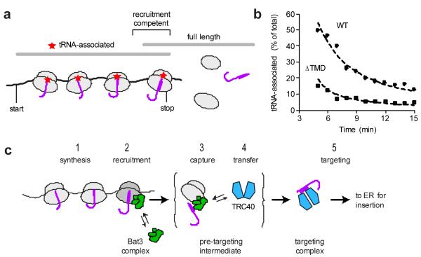
Hundreds of proteins are inserted post-translationally into the endoplasmic reticulum (ER) membrane by a single carboxy-terminal transmembrane domain (TMD). During targeting through the cytosol, the hydrophobic TMD of these tail-anchored (TA) proteins requires constant chaperoning to prevent aggregation or inappropriate interactions. A central component of this targeting system is TRC40, a conserved cytosolic factor that recognizes the TMD of TA proteins and delivers them to the ER for insertion. The mechanism that permits TRC40 to find and capture its TA protein cargos effectively in a highly crowded cytosol is unknown. Here we identify a conserved three-protein complex composed of Bat3, TRC35 and Ubl4A that facilitates TA protein capture by TRC40. This Bat3 complex is recruited to ribosomes synthesizing membrane proteins, interacts with the TMDs of newly released TA proteins, and transfers them to TRC40 for targeting. Depletion of the Bat3 complex allows non-TRC40 factors to compete for TA proteins, explaining their mislocalization in the analogous yeast deletion strains. Thus, the Bat3 complex acts as a TMD-selective chaperone that effectively channels TA proteins to the TRC40 insertion pathway.


Figures

Fig. 1
Fig. 1. Identification of a TMD-interacting protein complex
a, Cytosolic proteins bound and eluted from anti-Bat3 or anti-GFP (control) affinity columns are shown. HC and LC are IgG heavy and light chain. b, Sec61β (WT), a deletion construct lacking its TMD (ΔTMD), or the insertion-deficient 3R mutant were translated in reticulocyte lysate, affinity purified on an anti-Sec61β column, and immunoblotted for the indicated products. Total lysate was included for comparison. An autoradiograph of the blot revealed equal recovery of the three translated substrates. c, Crosslinking products of Sec61β (from pooled sucrose gradient fractions 6-9 in Sup. Fig. S4) were immunoprecipitated under denaturing or native conditions. Non-immune serum (N.I.S) was included as a control. d, Versions of Sec61β containing the TMD from the indicated proteins were analyzed for interaction with Bat3 and TRC40 by in vitro translation, crosslinking, and immunoprecipitation. An aliquot of the total translation reaction is shown for each substrate (‘subst.’), as well as the immunoprecipitation products of the crosslinking reactions. e, The TMD of Sec61β was mutated to change its hydrophobicity as indicated. f, Each construct was analyzed for its interactions with Bat3 complex and TRC40 as in panel b. An aliquot of the total translation product was analyzed by autoradiography to visualize the substrates.
Fig. 2
Fig. 2. The Bat3 complex mediates substrate capture by TRC40
a, Translation extracts were passed over anti-GFP (control), anti-Bat3, or anti-TRC40 affinity resins and different amounts of each depleted lysate analyzed by immunoblotting. b, Substrate capture assay using either total cytosol, or cytosol immunodepleted (‘Δ’) with the indicated affinity resins. In this assay (see Sup. Fig. S1), radiolabeled Sec61β RNCs are released with puromycin and capture by TRC40 is assessed by crosslinking. The portion of gel showing the TRC40-Sec61β crosslink is shown. Failure of TRC40 to capture substrate typically results in capture by a 38 kD protein (p38). The ‘addback’ lanes are Bat3-depleted cytosol replenished with affinity purified Bat3 complex (prepared using an anti-TRC35 resin; Sup. Fig. S8) at three concentrations spanning that present in the original cytosol. The ‘mock’ addback sample was prepared in parallel, but employed an irrelevant affinity resin (anti-GFP) in lieu of TRC35 affinity resin (Sup. Fig. S8). A reaction lacking crosslinker (XL) is shown in the first lane. Aliquots of each reaction (prior to crosslinking) were also analyzed by immunoblot against Bat3 and TRC40 to document their relative amounts in the reactions.
Fig. 3
Fig. 3. Bat3 complex captures substrates on ribosomes for transfer to TRC40
a, Ribosomes were purified under native conditions from reticulocyte lysate and analyzed for TRC40, Bat3, Ubl4A, and SRP54 by immunoblotting. Approximately 30-50% of SRP, 2-5% of Bat3 complex, and undetectable (<1%) amounts of TRC40 are ribosome-bound. b, Affinity and size purified RNCs (from 60 ul translation reactions) of Sec61β, a TMD-lacking version (ΔTMD), and the 3R mutant were analyzed by immunoblotting for the indicated antigens. For comparison, 0.5 ul translation lysate was analyzed. L9 is a ribosomal protein. Autoradiography confirmed equal recovery of each substrate. c, The amounts of ribosomes, Ubl4A, and SRP54 were quantified in total lysate, purified empty ribosomes, or purified RNC preparations. Shown are the amounts of SRP54 and Ubl4A, normalized to 100 ribosomes, averaged from multiple independent purifications (n=3 for empty ribosomes, n=7 for Sec61β-RNCs, and n=6 from β(3R)-RNCs). d, Translations of Sec61β in TRC40-depleted lysates lacking or replenished with recombinant zebrafish TRC40 were rapidly diluted, crosslinked, and separated by centrifugation into soluble and ribosome fractions. Sec61β, Bat3 crosslinks (immunopreciptiated with anti-Bat3), and TRC40 crosslinks are shown in the total (T), soluble (S), and ribosome (P) fractions. e, Cytosolic fractions from HT1080 cells lacking or stably over-expressing HA-tagged human TRC40 were bound to anti-HA resin and selectively eluted with TEV protease (which cleaves between the HA tag and TRC40). The eluted products, along with starting lysates, were analyzed by immunoblotting for Bat3, TRC40, or SGTA (a control protein). Note that endogenous TRC40 is present, but not visible at this exposure of the blot. Identical results were obtained when elution of the affinity column was with HA peptide instead of TEV protease (data not shown).
Fig. 4
Fig. 4. Translation termination is delayed for a TA protein
a, Schematic of Sec61β synthesis illustrating that nascent chains competent for recruitment of Bat3 complex would be near full-length and contain a covalently associated tRNA (red star). Such recruitment-competent polypeptides should migrate on gels close to the full-length size, but be precipitated by CTAB, which selectively precipitates tRNA-associated proteins. b, Time course of Sec61β and Sec61β(ΔTMD) (both tagged at the C-terminus with the same 12-residue epitope tag) synthesized at 25°C in reticulocyte lysate. At each time point, samples were analyzed directly or after CTAB precipitation. The proportion of total full-length polypeptide that was CTAB precipitated is plotted. The dashed lines indicate theoretical expectations (Sup. Fig. S15) for a t1/2 of translational termination of 15 and 60 sec. c, Model for shuttling of TA proteins from the ribosome to TRC40 via a pre-targeting intermediate involving recruitment of the Bat3 complex to the ribosome.

References

    1. High S, Gorlich D, Wiedmann M, Rapoport TA, Dobberstein B. The identification of proteins in the proximity of signal-anchor sequences during their targeting to and insertion into the membrane of the ER. J Cell Biol. 1991;113:35–44. - PMC - PubMed
    1. Fons RD, Bogert BA, Hegde RS. Substrate-specific function of the translocon-associated protein complex during translocation across the ER membrane. J. Cell Biol. 2003;160:529–39. - PMC - PubMed
    1. Rabu C, Schmid V, Schwappach B, High S. Biogenesis of tail-anchored proteins: the beginning for the end? J Cell Sci. 2009;122:3605–12. - PMC - PubMed
    1. Stefanovic S, Hegde RS. Identification of a targeting factor for posttranslational membrane protein insertion into the ER. Cell. 2007;128:1147–59. - PubMed
    1. Schuldiner M, et al. The GET complex mediates insertion of tail-anchored proteins into the ER membrane. Cell. 2008;134:634–45. - PMC - PubMed

Publication types

MeSH terms