. 2010 Sep;11(9):836-45.
doi: 10.1038/ni.1914. Epub 2010 Aug 1.

The transmembrane activator TACI triggers immunoglobulin class switching by activating B cells through the adaptor MyD88

Affiliations

The transmembrane activator TACI triggers immunoglobulin class switching by activating B cells through the adaptor MyD88

Bing He et al. Nat Immunol. 2010 Sep.

Abstract

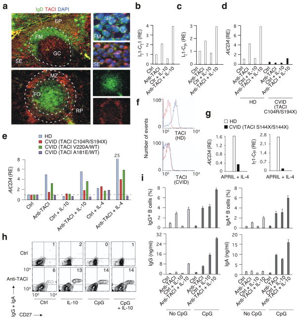
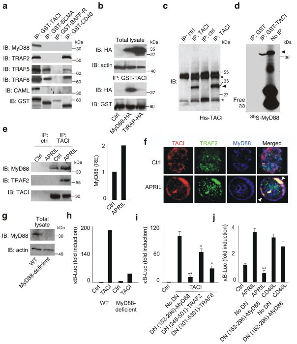
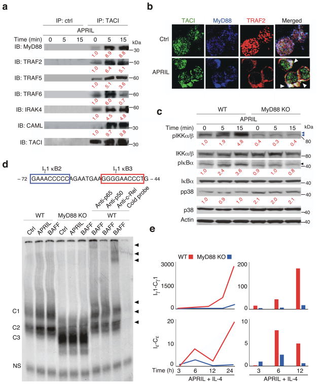
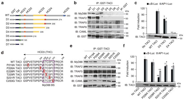
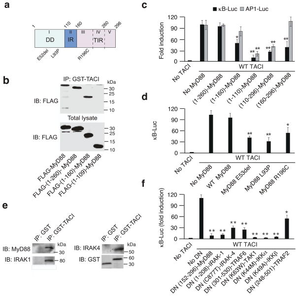
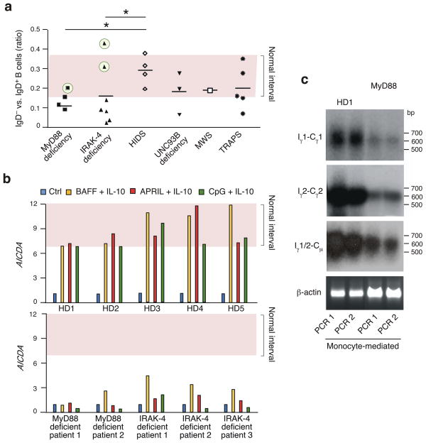
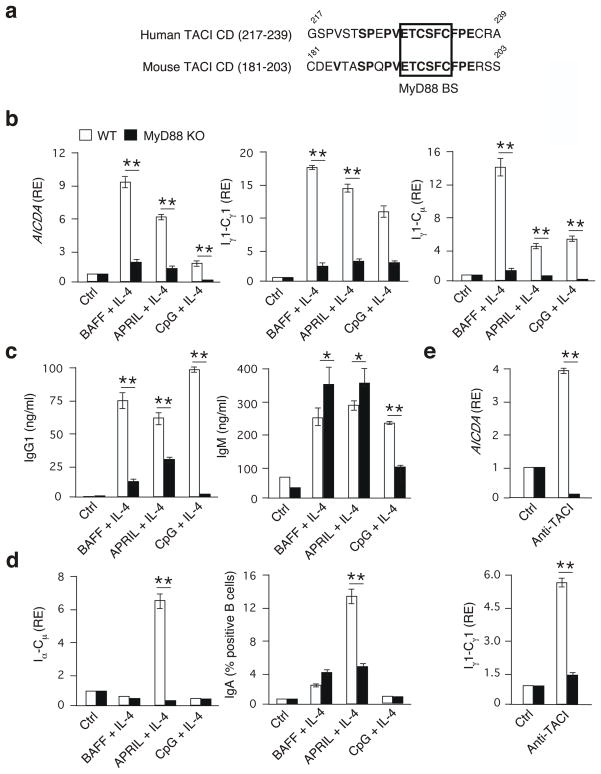
BAFF and APRIL are innate immune mediators that trigger immunoglobulin G (IgG) and IgA class-switch recombination (CSR) in B cells by engaging the receptor TACI. The mechanism that underlies CSR signaling by TACI remains unknown. Here we found that the cytoplasmic domain of TACI encompasses a conserved motif that bound MyD88, an adaptor that activates transcription factor NF-kappaB signaling pathways via a Toll-interleukin 1 (IL-1) receptor (TIR) domain. TACI lacks a TIR domain, yet triggered CSR via the DNA-editing enzyme AID by activating NF-kappaB through a Toll-like receptor (TLR)-like MyD88-IRAK1-IRAK4-TRAF6-TAK1 pathway. TACI-induced CSR was impaired in mice and humans lacking MyD88 or the kinase IRAK4, which indicates that MyD88 controls a B cell-intrinsic, TIR-independent, TACI-dependent pathway for immunoglobulin diversification.


Conflict of interest statement

COMPETING INTEREST STATEMENT

The authors declare that they have no competing financial interests.

Figures

Figure 1
Figure 1. TACI triggers CSR by cooperating with TLR ligands
(a) Immunofluorescence staining of tonsil (top) and splenic (bottom) tissues for IgD (green), TACI (red), and nuclei (blue). Dashed line, follicle; EP, epithelium; FM, follicular mantle; FO, follicle; GC, germinal center; MZ; marginal zone; SE, sub-epithelium; RP, red pulp. Original magnification, ×10 (left panels) or ×63 (right panels). (b–e) QRT-PCR of Iγ1-Cγ1, Iγ1-Cμ and AICDA in naïve (b–d) or lymphoblastoid (e) B cells from healthy donors (HD) or CVID patients with various heterozygous TACI substitutions cultured for 2 or 4 days with or without anti-TACI, IL-10 and/or IL-4. Results are normalized to ACTB (encoding β-actin) mRNA; RE, relative expression compared to B cells incubated with a control antibody (ctrl). (f) Flow cytometry of TACI on primary CD19+CD27+ B cells from a HD or CVID patient with homozygous S144X/S144X TACI substitution. Red histograms, ctrl; blue histograms, anti-TACI. (g) AICDA and Iγ1-Cμ in primary naive B cells from CVID case shown in f incubated with BAFF or APRIL plus IL-4 for 6 d. (h) Flow cytometry of IgG, IgA and CD27 on primary naive B cells incubated for 7 days with ctr, anti-TACI, IL-10 and/or CpG DNA. Numbers indicate percentages. (i) Flow cytometry of IgG and IgA (upper panels) and ELISA of secreted IgG and IgA (bottom panels) from B cells stimulated as in h. *P < 0.05 (one-tailed unpaired Student’s t-test). Data are from one of three experiments with similar results (a–h) or summarize three experiments (i; error bars, s.d.).
Figure 2
Figure 2. TACI interacts with MyD88
(a) GST-TACI, GST-BCMA, GST-BAFF-R or GST-CD40 immunoprecipitation (IP) of 2E2 B cell lysates, followed by immunoblotting (IB) of MyD88, TRAF2, TRAF5, TRAF6, CAML, or GST. kDa, kilodaltons. (b) Upper gel: IB of HA or actin in total lysates from 293 cells expressing control plasmid (ctrl), MyD88-HA or TIRAP-HA. Bottom gel: GST-TACI IP of 293 cell lysates, followed by IB of HA or GST. (c) Anti-TACI or anti-His IP of lysates from 2E2 B cells or TACI-His-expressing 293 cells, followed by IB for MyD88 or TACI. Asterisks, heavy (upper) and light (lower) chains of IP antibody; arrowhead, MyD88. (d) GST or GST-TACI IP of 35S-MyD88. Rightmost lane, 35S-MyD88 before IP; arrow, MyD88; free aa, free amino acids. (e) Anti-TACI or control antibody (ctrl) IP of lysates from human primary naïve B cells cultured for 15 min with medium (ctrl) or APRIL, followed by IB of MyD88, TRAF2 and TACI. Bars show intensity of MyD88 band relative to TACI in unstimulated B cells. (f) Confocal microscopy of TACI (red), TRAF2 (green) and MyD88 (blue) in primary naïve B cells exposed to medium (ctrl) or APRIL for 15 min. Arrowheads indicate co-localization. (g) IB of MyD88 and actin (loading control) from WT or MyD88-deficient 293 cells. (h,i) NF-κB reporter assay in WT or MyD88-deficient 293 cells transfected with TACI or a control empty plasmid (ctrl) in the presence or absence of DN-MyD88, DN-TRAF2 or DN-TRAF6. (j) NF-κB reporter assay in 2E2 B cells transfected with DN-MyD88 or a control empty plasmid (no DN) and incubated with medium (ctrl), APRIL or CD40L for 2 d. *P < 0.05 and ** P < 0.005 (one-tailed unpaired Student’s t-test). The data shown are from one of three experiments giving similar results (a–h) or summarize three experiments (i,j; error bars, SEM).
Figure 3
Figure 3. TACI requires MyD88 to activate NF-κB
(a) Immunoblotting (IB) of MyD88, TRAF2, TRAF5, TRAF6, IRAK-4, CAML and TACI upon control (ctrl) antibody or anti-TACI immunoprecipitation (IP) of lysates from primary mouse B cells cultured for 0, 5 or 15 min with APRIL. Red numbers indicate band intensity relative to TACI in unstimulated B cells. kDa, kilodaltons. (b) Confocal microscopy of TACI (green), TRAF2 (red) and MyD88 (blue) in primary mouse B cells exposed to medium (ctrl) or APRIL for 15 min. Arrowheads point to co-localization. (c) IB of phosphorylated (p) IKKα/β, IKKα/β, pIκBα, pIκBα, pp38, p38 and actin in primary WT or MyD88 KO B cells incubated with APRIL for 0, 5 or 15 min. Blue arrowheads point to pIKKβ (upper) and pIKKα (lower); black arrowhead points to pIκBα. Red numbers indicate band intensity relative to IKKα/β, IκBα and p38 in unstimulated B cells. (d) EMSA of NF-κB binding to the Iγ1 promoter in mouse primary B cells cultured with medium (ctrl), BAFF or APRIL for 3 h. Numbers indicate nucleotide positions compared to transcription initiation site. C1-C3, specific protein-DNA complexes; black arrowheads, supershifts; NS, non-specific band. (e) QRT-PCR of Iγ1-Cγ1 and Iε-Cε transcripts in primary WT (red) or MyD88 KO (blue) naïve B cells incubated with APRIL and IL-4 for various time points. Results are normalized to ACTB (encoding β-actin) mRNA; RE, relative expression compared to unstimulated B cells. Curves, time course; bars, earliest time points. Data are from one of three experiments giving similar results.
Figure 4
Figure 4. TACI binds MyD88 through a THC domain
(a) Scheme of WT TACI and D1-D7 GST-TACI deletion mutants. Numbers, carboxy-terminal residues; TD, transmembrane domain; HCD, highly conserved domain. Complete TACI sequence shown in Supplementary Fig. S8. (b) Immunoprecipitation (IP) of 2E2 B cell lysates with GST, WT GST-TACI, or GST-TACI deletion mutants followed by immunoblotting (IB) of MyD88, TRAF2, TRAF5, TRAF6, CAML, or GST. kD, kilodaltons. (c) NF-κB and AP1 reporter assays in 293 cells expressing no TACI, WT TACI, D3 TACI or D4 TACI. Bottom gel, IB of TACI from total lysates. (d) Site-directed mutations in the THC domain. Upper numbers, residue positions; red crosses, substitutions; purple box, MyD88-binding site (BS). (e) IP of 2E2 B cell lysates with WT GST-TACI or GST-TACI site-directed mutants followed by IB of MyD88, TRAF2, TRAF5, TRAF6, CAML, or GST. (f) NF-κB and AP1 reporter assays in 293 cells expressing no TACI, WT TACI, or TACI site-directed mutants. Bottom gel, IB of TACI from total cell lysates. *P < 0.05 and ** P < 0.005 (one-tailed unpaired Student’s t-test). The data shown are from one of three experiments giving similar results (c,f) or summarize three experiments (d,g; error bars, SEM).
Figure 5
Figure 5. TACI signals through a TLR-like pathway
(a) Structure of MyD88; top Arabic numerals, residues delimiting DD, IR and TIR; Roman numerals (I to V), exons; bottom codes, mutations causing MyD88 deficiency. (b) Immunoblotting (IB) of FLAG from 293 cells expressing wt FLAG-MyD88 or FLAG-MyD88 deletion mutants before (total lysate) or after immunoprecipitation (IP) with GST-TACI. (c,d) NF-κB and AP1 reporter assays in 293 cells expressing wt MyD88 or various deletion and site-directed MyD88 mutant proteins in the presence or absence of TACI. *P < 0.05 or **P < 0.005 versus no MyD88 (one-tailed unpaired Student’s t-test). (e) IP of 2E2 B cell lysates with GST or GST-TACI, followed by IB of MyD88, IRAK-1, IRAK-4 or GST (loading control). (f) NF-κB reporter assays in 293 cells with and without wt TACI expression in the presence or absence of DN-MyD88, DN-IRAK-1, DN-IRAK-4, DN-TRAF6, DN-TAK1, DN-IKKα, DN-IKKβ, or DN-TRAF2. *P < 0.05 and ** P < 0.005, versus no DN (one-tailed unpaired Student’s t-test). Data represent one of three experiments giving similar results (b,e) or summarize three experiments (c,d,f; error bars, SEM).
Figure 6
Figure 6. TACI requires MyD88 to induce CSR in humans
(a) Flow cytometric analysis of IgD/IgD+ B-cell ratio in the peripheral blood of patients with MyD88 or IRAK-4 deficiency and control age-matched patients with hyper-IgD syndrome (HIDS), UNC93B deficiency, Mukle-Wells syndrome (MWS) or TNF receptor-associated periodic fever syndrome (TRAPS). Pink area delimits range of IgD/IgD+ B-cell ratio in healthy donors (normal interval). Circled cases are further studied in Supplementary Fig. S12. *P <0.05 (two-tailed paired Student’s t-test). (b) QRT-PCR of AICDA transcripts in B cells from healthy donors (HD) and patients with MyD88 or IRAK-4 deficiency, after culture for 4 d in the presence or absence of BAFF plus IL-10, APRIL plus IL-10 or CpG DNA plus IL-10. Results are normalized to ACTB (encoding β-actin) mRNA; RE, relative expression compared to control (ctrl) unstimulated B cells. (c) Southern blot analysis of RT-PCR-amplified Iγ1-Cγ1, Iγ2-Cγ2, and Iγ1/2-Cμ transcripts in B cells from healthy or MyD88-deficient subjects exposed to BAFF- and APRIL-expressing monocytes. Additional controls shown in Supplementary Fig. S13. PCR1 and PCR2 indicate independent PCR amplifications; bp, base pairs.
Figure 7
Figure 7. TACI requires MyD88 to induce CSR in mice
(a) MyD88-binding site (BS) in human or mouse TACI. Numbers, amino-acid positions; bold letters, identical amino acids; box, MyD88-binding site in the THC domain. (b) QRT-PCR of AICDA, Iγ1-Cγ1 and Iγ1-Cμ transcripts from WT (open bars) or MyD88 KO (solid bars) mouse B cells cultured for 4 d in the presence or absence of BAFF, APRIL or CpG DNA plus IL-4. Results are normalized to ACTB (encoding β-actin) mRNA; RE, relative expression compared to control (ctrl) unstimulated B cells. (c) ELISA of IgG1 and IgM from WT or MyD88 KO B cells cultured as in c for 8 days. (d,e) QRT-PCR of Iα1-Cμ and flow cytometric analysis of surface IgA from WT or MyD88 KO B cells cultured as in b for 48 h (Iα1-Cμ) or 5 days (IgA). (e) QRT-PCR of AICDA and Iγ1-Cγ1 from WT or MyD88 KO B cells cultured with a ctrl antibody or anti-TACI for 2 d. *P < 0.05, versus wild type (one-tailed unpaired Student’s t-test). The data presented summarize three independent experiments performed by pooling splenic naive B cells from three mice (error bars, SEM).

References

    1. Cooper MD, Alder MN. The evolution of adaptive immune systems. Cell. 2006;124:815–822. - PubMed
    1. Schlissel MS. Regulating antigen-receptor gene assembly. Nat Rev Immunol. 2003;3:890–899. - PubMed
    1. Stavnezer J, Guikema JE, Schrader CE. Mechanism and regulation of class switch recombination. Annu Rev Immunol. 2008;26:261–292. - PMC - PubMed
    1. Muramatsu M, et al. Class switch recombination and hypermutation require activation-induced cytidine deaminase (AID), a potential RNA editing enzyme. Cell. 2000;102:553–563. - PubMed
    1. Cerutti A. The regulation of IgA class switching. Nat Rev Immunol. 2008;8:421–434. - PMC - PubMed

Publication types

MeSH terms

Substances