. 2010 Aug;16(8):909-14.
doi: 10.1038/nm.2186. Epub 2010 Aug 1.

MicroRNA-132-mediated loss of p120RasGAP activates the endothelium to facilitate pathological angiogenesis

Affiliations

MicroRNA-132-mediated loss of p120RasGAP activates the endothelium to facilitate pathological angiogenesis

Sudarshan Anand et al. Nat Med. 2010 Aug.

Abstract

Although it is well established that tumors initiate an angiogenic switch, the molecular basis of this process remains incompletely understood. Here we show that the miRNA miR-132 acts as an angiogenic switch by targeting p120RasGAP in the endothelium and thereby inducing neovascularization. We identified miR-132 as a highly upregulated miRNA in a human embryonic stem cell model of vasculogenesis and found that miR-132 was highly expressed in the endothelium of human tumors and hemangiomas but was undetectable in normal endothelium. Ectopic expression of miR-132 in endothelial cells in vitro increased their proliferation and tube-forming capacity, whereas intraocular injection of an antagomir targeting miR-132, anti-miR-132, reduced postnatal retinal vascular development in mice. Among the top-ranking predicted targets of miR-132 was p120RasGAP, which we found to be expressed in normal but not tumor endothelium. Endothelial expression of miR-132 suppressed p120RasGAP expression and increased Ras activity, whereas a miRNA-resistant version of p120RasGAP reversed the vascular response induced by miR-132. Notably, administration of anti-miR-132 inhibited angiogenesis in wild-type mice but not in mice with an inducible deletion of Rasa1 (encoding p120RasGAP). Finally, vessel-targeted nanoparticle delivery of anti-miR-132 restored p120RasGAP expression in the tumor endothelium, suppressed angiogenesis and decreased tumor burden in an orthotopic xenograft mouse model of human breast carcinoma. We conclude that miR-132 acts as an angiogenic switch by suppressing endothelial p120RasGAP expression, leading to Ras activation and the induction of neovascularization, whereas the application of anti-miR-132 inhibits neovascularization by maintaining vessels in the resting state.


Figures

Figure 1
Figure 1
miR-132 regulates growth factor–mediated angiogenesis in vitro and in vivo. (a) HUVEC cell proliferation. After transfection with miR-132 or anti–miR-132 or a control (Ctrl) miRNA, HUVECs were pulsed with BrdU and cell proliferation was measured using an ELISA assay. One representative experiment of three is shown, with the average values of triplicate wells. *P < 0.01 compared to control miRNA. (b) HUVEC tube formation. 24 h after transfection as in a, HUVECs were suspended in a three-dimensional collagen matrix. Tube lengths were measured using MetaMorph software on day 4. One representative experiment of three is shown, with the average values of triplicate wells. *P < 0.01 compared to control miRNA. (c) Angiogenesis in Matrigel plugs in vivo. Growth factor–reduced Matrigel containing either PBS or bFGF was injected subcutaneously into C57BL/6 mice. Mice received 10 µg of either a control anti-miRNA or anti–miR-132 in PBS intravenously (n = 6 per group). Angiogenesis was quantified by measuring FITC-lectin content on day 5. *P < 0.05 for control bFGF plugs compared to anti–miR-132 bFGF plugs. Right micrographs show representative Matrigel plugs from each group. Scale bar, 1 cm. (d) Retinal angiogenesis. Either control anti-miRNA or anti–miR-132 (1 µg) was injected intraocularly into 6-d-old BALB/c pups (n = 5 per group). Retinas were collected and stained with CD31-specific monoclonal antibodies (mAb). Bars show mean CD31 area (n = 25 fields) calculated using MetaMorph software. *P < 0.01. Right micrographs show representative confocal images of the deep plexus retinal vasculature. White dashed lines indicate the periphery of the retinas. Scale bar, 100 µm. Bars show means ± s.e.m.
Figure 2
Figure 2
Endothelial activation mediated by miR-132 depends on its downregulation of p120RasGAP. (a) Luciferase activity of 293T cells co-transfected with a luciferase-Rasa1 3′ UTR plasmid containing either the WT 3′ UTR or a mutated sequence, a β-galactosidase plasmid and either a control miRNA or miR-132. Bars shown mean relative luciferase activity normalized to the β-galactosidase levels (n = 3). *P < 0.05 compared to control miRNA. (b) Western blot detecting p120RasGAP in HUVECs transfected with either a control miRNA or miR-132. One representative experiment of two is shown. (c) Ras activity in HUVECs transfected with control miRNA, miR-132, control anti-miRNA or anti–miR-132. One representative experiment of two is shown, with the average value of triplicate wells. *P < 0.05 compared to control miRNA or control anti-miRNA. (d) HUVEC cell proliferation. After transfection with two different siRNAs (1 and 2) targeting p120RasGAP or with miR-132, HUVECs were pulsed with BrdU and cell proliferation was measured using an ELISA assay. Ctrl, control siRNA. One representative experiment of three is shown. *P < 0.05. Cell proliferation (e) and tube formation (f) of HUVECs. Cells were transfected with a control vector or a miRNA-resistant form of Rasa1 and, 24 h later, were transfected with miR-132 or a control miRNA. The values shown are for miR-132 transfection relative to control miRNA transfection. One representative experiment of two is shown. P < 0.05 (miR-132 compared with control miRNA). (g) Angiogenesis in Matrigel plugs in vivo. Forty-eight hours after mice were injected with bFGF Matrigel plugs, they were injected with either a control plasmid (vector) or a plasmid encoding miRNA-resistant Rasa1 in nanoparticles. After a further 24 h, mice in each group (n = 3 per group) were treated with either a control miRNA or miR-132 in RGD nanoparticles. Angiogenesis in the plugs was quantified by measuring FITC-lectin content. The values shown are for mice receiving miR-132 normalized to those receiving control miRNA. One representative experiment of two is shown. P < 0.05 (miR-132 compared with control miRNA). (h) Angiogenesis in Matrigel plugs in vivo. Tamoxifen-treated Rasa1fl/fl Ert2-ubiquitin-Cre mice (Rasa1−/−) (n = 3 per group) and wild-type mice (n = 3 per group) were injected with bFGF-containing Matrigel plugs and 2 h later treated with either a control anti-miRNA (Ctrl) or anti–miR-132. Angiogenesis was quantified by measuring FITC-lectin content. *P < 0.05 (anti–miR-132 compared with control anti-miRNA in WT mice). Bars show means ± s.e.m.
Figure 3
Figure 3
miR-132 and p120RasGAP are expressed reciprocally in quiescent versus proliferative endothelium. (a,b) Normal human breast tissue or human breast carcinoma was analyzed for CD31 and p120RasGAP expression by immunofluorescence (a) and with miR-132 expression by in situ hybridization (b). Quantification of the results for 12 breast carcinoma tissue sections and adjacent normal tissue is also shown. Arrows and asterisks indicate blood vessels in the normal and tumor sections. Scale bar, 100 µm. *P < 0.05. (c) In situ hybridization for miR-132 and staining for p120RasGAP was performed on a human hemangioma tissue array. Representative H&E sections and miR-132 staining is shown for hemangiomas from mesentery and brain. Quantification is shown for miR-132 expression as the fold increase over normal tissue and for RasGAP expression scored on a 1–4 scale. miR-132 expression was quantified using MetaMorph software. Scale bar, 100 µm. n = 68 hemangioma samples; n = 32 normal samples. *P < 0.05, Mann-Whitney U test. Bars show means ± s.e.m.
Figure 4
Figure 4
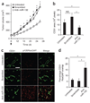
Targeted delivery of anti–miR-132 decreases tumor burden by restoring endothelial p120RasGAP. Nude mice were implanted with 2 × 10⁶ MDA-MB 231 cells in the mammary fat pad. The tumors were measured 8 d later, and mice with palpable tumors of similar volumes were randomly assigned to three groups. Mice were left untreated or received 50 µg of scrambled anti-miRNA or anti–miR-132 every 48 h until day 24. (a) Tumor volumes (mean ± s.e.m) of at least seven mice per group, starting from the day of treatment (black arrow). *P < 0.05 compared to anti–miR-132. (b–d) Immunofluorescent staining of CD31 and p120RasGAP in tumor sections from at least three mice per group. (b) Microvessel density measured by CD31 area. (c) Representative images of p120RasGAP expression in CD31-positive cells. Scale bar, 100 µm. (d) Quantification of the overlap of CD31 and p120RasGAP expression (percentage of CD31 cells that are also positive for RasGAP expression). Pixel density was quantified using MetaMorph software. Bars are mean ± s.e.m of multiple images from at least three mice per group. *P < 0.05 (anti–miR-132 compared to untreated or scrambled). NS, not significant.

Comment in

Similar articles

Cited by

References

    1. Murphy EA, et al. Nanoparticle-mediated drug delivery to tumor vasculature suppresses metastasis. Proc. Natl. Acad. Sci. USA. 2008;105:9343–9348. - PMC - PubMed
    1. Hobson B, Denekamp J. Endothelial proliferation in tumours and normal tissues: continuous labelling studies. Br. J. Cancer. 1984;49:405–413. - PMC - PubMed
    1. Folkman J. The role of angiogenesis in tumor growth. Semin. Cancer Biol. 1992;3:65–71. - PubMed
    1. Fish JE, Srivastava D. MicroRNAs: opening a new vein in angiogenesis research. Sci. Signal. 2009;2:pe1. - PMC - PubMed
    1. Lindquist JN, Cheresh DA, Snyder EY. Derivation of vasculature from embryonic stem cells. Curr. Protoc. Stem Cell Biol. 2010;12:1.1F.9.1–1.1F.9.6. - PubMed

Publication types

MeSH terms