. 2010 Sep;12(5):677-89.
doi: 10.1038/aja.2010.56. Epub 2010 Aug 2.

Growth inhibition mediated by PSP94 or CRISP-3 is prostate cancer cell line specific

Affiliations

Growth inhibition mediated by PSP94 or CRISP-3 is prostate cancer cell line specific

Bhakti R Pathak et al. Asian J Androl. 2010 Sep.

Abstract

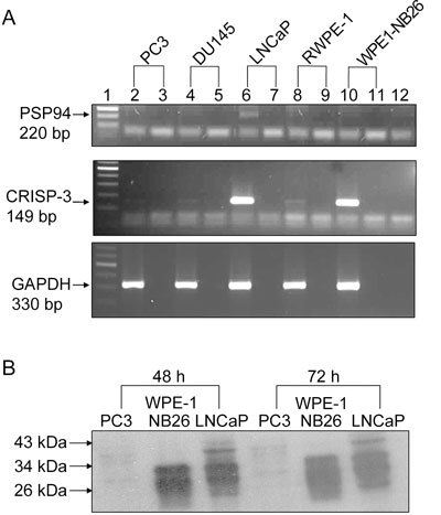
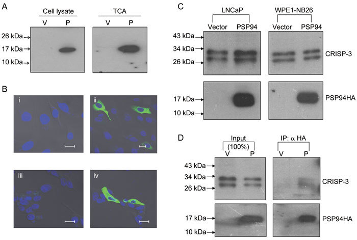
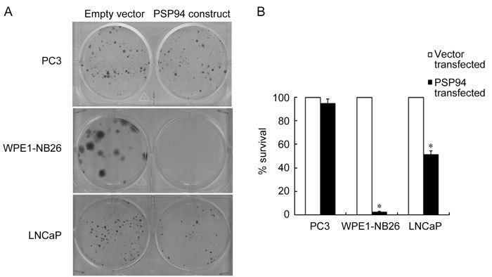
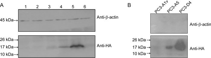
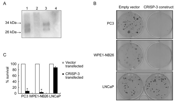
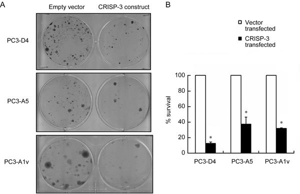
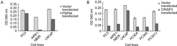
The prostate secretory protein of 94 amino acids (PSP94) has been shown to interact with cysteine-rich secretory protein 3 (CRISP-3) in human seminal plasma. Interestingly, PSP94 expression is reduced or lost in the majority of the prostate tumours, whereas CRISP-3 expression is upregulated in prostate cancer compared with normal prostate tissue. To obtain a better understanding of the individual roles these proteins have in prostate tumourigenesis and the functional relevance of their interaction, we ectopically expressed either PSP94 or CRISP-3 alone or PSP94 along with CRISP-3 in three prostate cell lines (PC3, WPE1-NB26 and LNCaP) and performed growth inhibition assays. Reverse transcription-polymerase chain reaction and Western blot analysis were used to screen prostate cell lines for PSP94 and CRISP-3 expression. Mammalian expression constructs for human PSP94 and CRISP-3 were also generated and the expression, localization and secretion of recombinant protein were assayed by transfection followed by Western blot analysis and immunofluorescence assay. The effect that ectopic expression of PSP94 or CRISP-3 had on cell growth was studied by clonogenic survival assay following transfection. To evaluate the effects of co-expression of the two proteins, stable clones of PC3 that expressed PSP94 were generated. They were subsequently transfected with a CRISP-3 expression construct and subjected to clonogenic survival assay. Our results showed that PSP94 and CRISP-3 could each induce growth inhibition in a cell line specific manner. Although the growth of CRISP-3-positive cell lines was inhibited by PSP94, growth inhibition mediated by CRISP-3 was not affected by the presence or absence of PSP94. This suggests that CRISP-3 may participate in PSP94-independent activities during prostate tumourigenesis.


Figures

Figure 1
Figure 1
Pattern of endogenous PSP94 and CRISP-3 expression in different prostate cell lines. (A): Reverse transcription-polymerase chain reaction (RT-PCR) results of the total RNA content that was extracted from the five tested cell lines. The panels from the top to the bottom show amplification of PSP94, CRISP-3 and GAPDH, respectively. Lane 1 is a 100-bp ladder. Lanes 2, 4, 6, 8 and 10 are RT-PCR products for PC3, DU145, LNCaP, RWPE-1 and WPE1-NB26, respectively. Lanes 3, 5, 7, 9 and 11 are PCR reactions for no RT enzyme controls for the same samples. Lane 12 is a water control. (B): Semi-quantitative Western blot analysis for detection of CRISP-3 protein. TCA-precipitated proteins from conditioned medium that was harvested at 48 h (left side) and 72 h (right side) from 5 × 10⁵ PC3 cells, WPE1-NB26 cells and LNCaP cells were subjected to Western blot analysis using an anti-CRISP-3 polyclonal antibody. The presence of two bands at around 30 and 28 kDa in the WPE1-NB26 and LNCaP cells represent glycosylated and unglycosylated CRISP-3 protein, respectively.
Figure 2
Figure 2
Secreted recombinant PSP94 interacts with endogenous CRISP-3. (A): Western blot analysis for detection of PSP94HA. Cell lysate and TCA-precipitated proteins derived from the conditioned medium from PC3 cells that were transiently transfected with either empty vector (V) or a PSP94 expression construct (P) were probed with anti-HA antibody. (B): Localization of PSP94HA by immunofluorescence. PC3 cells that were transiently transfected with empty vector (i) and a PSP94 expression construct (ii) as well as LNCaP cells transiently transfected with empty vector (iii) and a PSP94 expression construct (iv) were first probed with polyclonal anti-HA antibody and then with FITC-labelled secondary antibody. Bars = 20 μm. (C): Effect of ectopically expressed PSP94 on endogenous CRISP-3. TCA-precipitated proteins from vector-transfected and PSP94 expression construct-transfected WPE1-NB26 and LNCaP cells were subjected to Western blot analysis 48 h after transfection. The upper panel was probed with anti-CRISP-3 antibody and the lower panel was probed with anti-HA antibody. (D): Co-immunoprecipitation of PSP94 and CRISP-3 from the conditioned medium of vector-transfected and PSP94 expression construct-transfected WPE1-NB26 cells. PSP94HA was immunoprecipitated using anti-HA agarose-conjugated beads. The upper portion of the Western blot was probed with anti-CRISP-3 antibody and the lower portion was probed with anti-HA antibody. The lanes marked with input show the total amount of CRISP-3 and PSP94HA that was immunoprecipitated from the conditioned medium.
Figure 3
Figure 3
Ectopic expression of PSP94 has an inhibitory effect on cell growth in a cell line specific manner. (A): A representative composite picture of the crystal violet staining from the clonogenic survival assay. A single well (of the six-well plate) is shown for each cell line (PC3, WPE1-NB26 and LNCaP) following transfection with empty vector, pcDNA3.1+ (left wells) or PSP94 expression construct (right wells). At 24 h after transfection, the cells were split and stable clones were selected on G418. Clones obtained after two weeks of selection were stained and counted. (B): Effect of the over-expression of PSP94 on the clonogenic survival of PC3, WPE1-NB26 and LNCaP cells. For each cell line, the number of clones obtained in the vector-transfected wells was considered to represent 100% survival. The number of clones obtained in the PSP94 expression construct-transfected wells was used to calculate the percentage of surviving cells with respect to the empty vector-transfected cells. The assay was repeated at least three times for each cell line. The graph is the average of three independent observations and is plotted as the mean percentage survival ± SE. The observed reduction in the percentage survival of WPE1-NB26 and LNCaP cells was statistically significant when compared with PC3 cells (*P < 0.05, compared with PC3 cells; one-way ANOVA).
Figure 4
Figure 4
Screening of stable clones expressing PSP94 by Western blot analysis. (A): Lanes 1–5 represent cell lysates from five different clones following PSP94 expression construct transfection of PC3 cells. Lane 6 is a cell lysate from one of the clones that was obtained following transfection with empty vector (pcDNA3.1+). The expression of recombinant PSP94HA was confirmed by probing the blot with anti-HA antibody. β-Actin was used as a loading control. (B): Western blot analysis of the TCA-precipitated proteins from the culture supernatants of stable clones PC3-A1v (which carried empty vector), PC3-A5 (which expressed PSP94HA) and PC3-D4 (which expressed PSP94HA) after roughly 30 passages in culture. Clones PC3-A5 and PC3-D4 stably expressed and secreted PSP94HA. The blot was probed with anti-HA antibody to detect PSP94HA, and with anti-β-actin antibody to show that the conditioned media were not contaminated with cellular contents.
Figure 5
Figure 5
Cell line specific growth-inhibitory effect caused by CRISP-3 over-expression. (A): Expression of recombinant CRISP-3 was detected by Western blot analysis using anti-CRISP-3 antibody. TCA-precipitated proteins from the conditioned medium of PC3 cells that were transiently transfected with CRISP-3 expression construct (Lane 1) showed the presence of glycosylated and unglycosylated CRISP-3 similar to the positive control (Lane 3) that contained TCA-precipitated proteins from the conditioned medium of WPE1-NB26 cells. TCA-precipitated proteins from the conditioned medium of PC3 cells transfected with empty vector (Lane 2) as well as untransfected RWPE-1 (Lane 4) served as a negative control. (B): A representative composite picture of the crystal violet staining from the clonogenic survival assay. A single well (of the six-well plate) is shown for each cell line (PC3, WPE1-NB26 and LNCaP) that was transfected with either the empty vector, pEFIRES-P (left wells), or the CRISP-3 expression construct (right wells). At 24 h after transfection, the cells were split and stable clones were selected on puromycin. Clones obtained after 2 weeks of selection were stained and counted. (C): Effect of over-expression of CRISP-3 on the clonogenic survival of PC3, WPE1-NB26 and LNCaP cells. The percentage survival with respect to empty vector was calculated as described above. The assay was repeated at least three times for each cell line. The graph is the average of three independent observations and is plotted as mean percentage survival ± SE. The reduction in percentage survival of PC3 and WPE1-NB26 cells was statistically significant when compared with LNCaP cells (*P < 0.05, compared with LNCaP; one-way ANOVA).
Figure 6
Figure 6
The growth-inhibitory effect of CRISP-3 is not modulated by PSP94. (A): A representative composite picture of the crystal violet staining from the clonogenic survival assay for the stable clones PC3-D4, PC3-A5 (PSP94-expressing clones) and PC3-A1v (empty vector-transfected clone). A single well (of the six-well plate) is shown for each clone that was transfected with either empty vector, pEFIRES-P (left wells), or the CRISP-3 expression construct (right wells). At 24 h after transfection, cells were split and selected on puromycin. Clones obtained after 2 weeks of selection were stained and counted. (B): The effect of over-expression of CRISP-3 on the clonogenic survival of PC3-D4, PC3-A5 and PC3-A1v. The percentage survival with respect to empty vector was calculated as described above. The assay was repeated at least three times for each cell line. The graph is the average of three independent observations and is plotted as the mean percentage survival ± SE. The reduction in percentage survival of all three clones was statistically significant when compared with LNCaP (*P < 0.05, compared with LNCaP shown in Figure 5B; one-way ANOVA).
Figure 1S
Figure 1S
Colorimetric assay for the clonogenic survival after transfection. Absorbance at 590 nm was measured for the cell associated dye from stable clones obtained after transfection with PSP94 expression construct and empty vector (A) and for those obtained after CRISP-3 transfection and empty vector transfection (B). Optical density is plotted as mean ± SE. The experiment was repeated thrice and the graph is for one representative experiment.

Comment in

Similar articles

Cited by

References

    1. Doctor VM, Sheth AR, Simha MM, Arbatti NJ, Aaveri JP, et al. Studies on immunocytochemical localization of inhibin-like material in human prostatic tissue: comparison of its distribution in normal, benign and malignant prostates. Br J Cancer. 1986;53:547–54. - PMC - PubMed
    1. Lilja H, Abrahamsson PA. Three predominant proteins secreted by the human prostate gland. Prostate. 1988;12:29–38. - PubMed
    1. Shukeir N, Garde S, Wu JJ, Panchal C, Rabbani SA. Prostate secretory protein of 94 amino acids (PSP-94) and its peptide (PCK3145) as potential therapeutic modalities for prostate cancer. Anti-Cancer Drugs. 2005;16:1045–51. - PubMed
    1. Garde SV, Basrur VS, Li L, Finkelman MA, Krishan A, et al. Prostate secretory protein (PSP94) suppresses the growth of androgen-independent prostate cancer cell line (PC3) and xenografts by inducing apoptosis. Prostate. 1999;38:118–25. - PubMed
    1. Cadieux PA, Mikolajczak SA, Reeves J, Strathdee C, Reid G, et al. Rat PSP94 inhibits the growth and viability of the rat adenocarcinoma cell line PAIII in vitro. Cancer Invest. 2006;24:242–51. - PubMed

Publication types

MeSH terms