. 2010 Oct 14;29(41):5559-67.
doi: 10.1038/onc.2010.326. Epub 2010 Aug 2.

p66(Shc) restrains Ras hyperactivation and suppresses metastatic behavior

Affiliations

p66(Shc) restrains Ras hyperactivation and suppresses metastatic behavior

Z Ma et al. Oncogene. .

Abstract

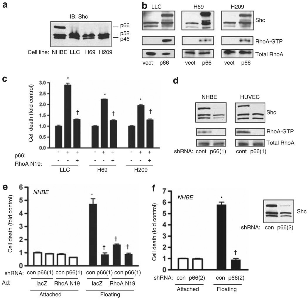
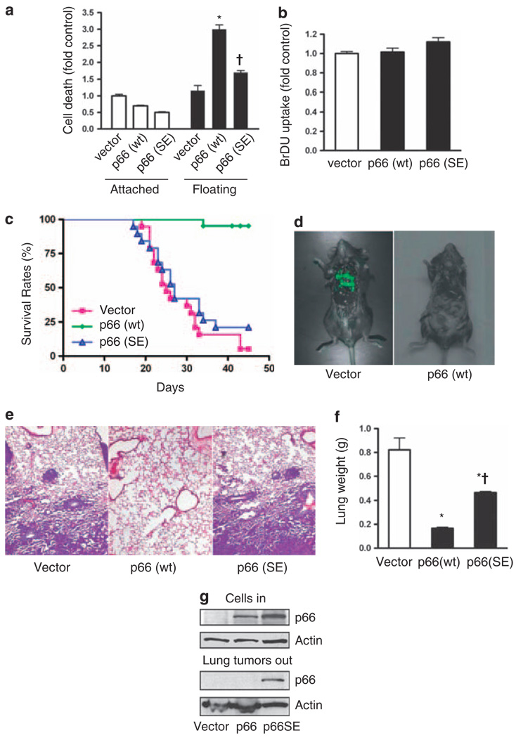
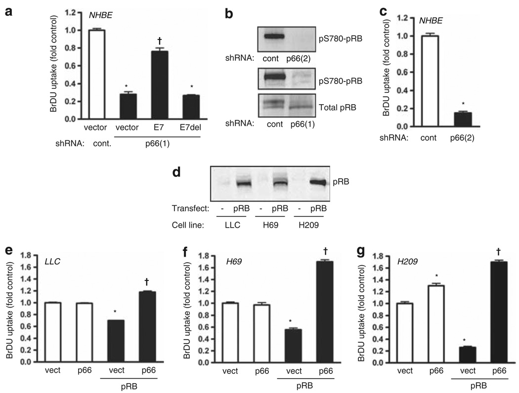
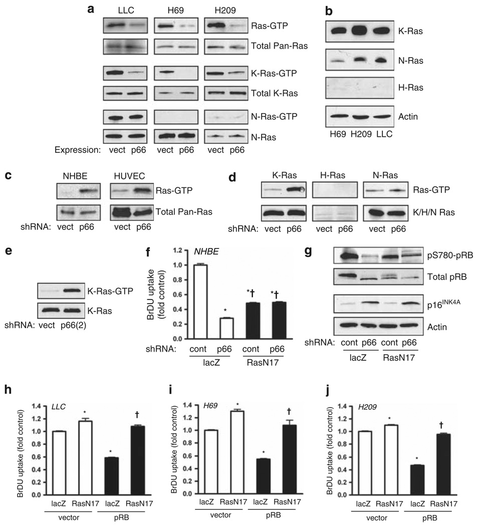
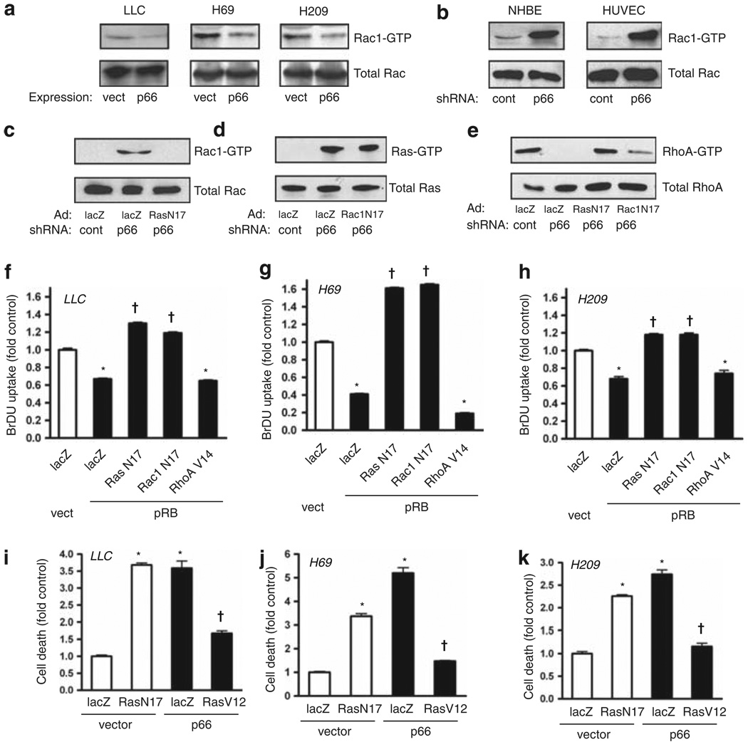
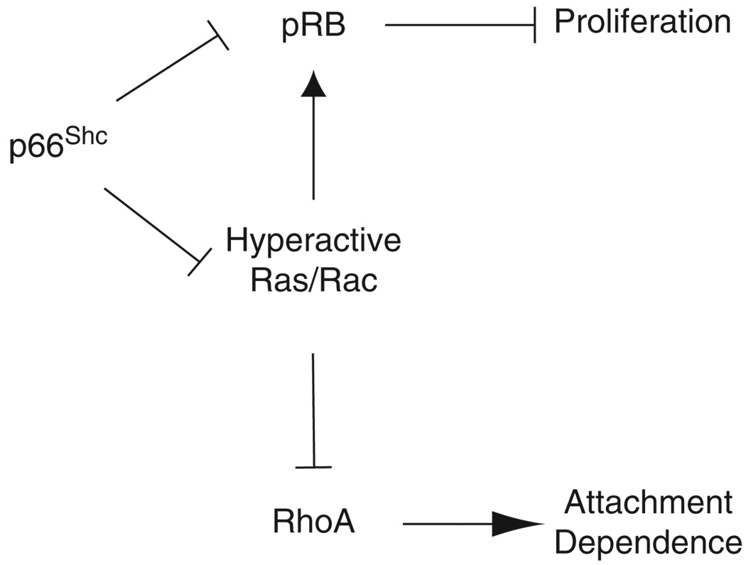
Normal tissue cells survive and proliferate only while anchored to solid substrate. Conversely, transformed cells both survive and proliferate following detachment, having lost attachment context through unclear mechanisms. p66(Shc) is a focal adhesion-associated protein that reports cell attachment through a RhoA-dependent mechanosensory test. We find that human small cell lung cancer (SCLC) cells and mouse Lewis lung carcinoma (LLC), which display aggressive metastatic behavior, lack both p66(Shc) and retinoblastoma (pRB) and bypass anoikis. Re-expression of p66(Shc) in these cells restores anoikis and provides striking protection from metastasis by LLC cells in vivo. Notably, knockdown of p66(Shc) in normal epithelial cells leads to unrestrained Ras activation, preventing anoikis through downstream suppression of RhoA but blocking proliferation in a pRB-dependent manner, thus mimicking oncogenic Ras. Conversely, LLC and SCLC cells display constitutive Ras activation necessary to bypass anoikis, which is reversed by re-expression of p66(Shc). p66(Shc) therefore coordinates Ras-dependent control of proliferation and anchorage sensation, which can be defeated in the evolution of highly metastatic tumors by combined loss of both p66(Shc) and pRB.


Conflict of interest statement

Conflict of interest

The authors declare no conflict of interest.

Figures

Figure 1
Figure 1
p66Shc promotes anoikis through RhoA. (a) Immunoblot for Shc demonstrates expression of three Shc isoforms by normal human bronchial epithelium (NHBE) cells but loss of p66Shc expression by Lewis lung carcinoma (LLC) and SCLC (H69 and H209) cell lines. (b) LLC, H69 and H209 cells were transduced with p66Shc. The top panels demonstrate Shc isoform expression. Active RhoA was assessed by pulldown and was shown to consistently increase following expression of p66Shc. (c) LLC, H69 and H209 cells were grown in low attachment plates after lentiviral transduction with p66Shc or empty vector, and adenoviral transduction with dominant-negative RhoA(N19) or lacZ (control). After 16 h, cell death was assessed. Mean ± s.e.m. of four determinations is shown. *P < 0.001 compared with respective control, P < 0.001 compared with p66Shc alone. (d) p66Shc was knocked down with shRNA in NHBE cells and human umbilical vein endothelial cells (HUVECs). The top panels demonstrate knockdown effect. The bottom panels show that p66Shc knockdown completely suppressed RhoA activity. (e) NHBE cells were transduced with the indicated shRNA by lentivirus and RhoA(N19) or lacZ by adenovirus, and allowed to adhere or forced to float for 16 h. Cell death was measured as above. Mean ± s.e.m. of four determinations is shown. *P < 0.001 compared with attached control, P < 0.001 compared with floating control. (f) NHBE cells were transduced with a second shRNA against p66Shc (shRNA(2)) and cell death measured as above. Inset shows selective effect of shRNA(2) for p66Shc. Mean ± s.e.m. of four determinations is shown. *P < 0.001 compared with attached control, P < 0.001 compared with floating control.
Figure 2
Figure 2
p66Shc suppresses lung metastasis in vivo. (a) LLC cells stably expressing p66Shc or p66(S36E) were plated on normal or low attachment plates for 16 h and cell death assessed. Expression of wild-type p66Shc increased detachment-induced cell death (*P < 0.001 from vector control), whereas p66(S36E) had a reduced effect (P < 0.001 from p66Shc floating). Mean ± s.e.m. of four determinations is shown. (b) 5-bromodeoxyuridine (BrDU) uptake of attached LLC cells expressing p66Shc (p66 wt) or p66(S36E) was not different from vector control. (c) LLC cells expressing p66Shc or p66(S36E)were injected into the tail vein of 6-week-old female C57BL/6 mice. Kaplan–Meier survival curve is shown; survival between groups was different, P < 0.0001. n = 19 (vector), n = 21 (p66 wt) and n = 19 (p66(SE)). (d) Representative mice receiving vector-LLC or p66-LLC cells. Multiple GFP-positive lung metastases are apparent in the former but not in the latter group. (e) Representative hematoxylin and eosin stain of lungs from mice injected with vector control, p66Shc or p66(S36E)-expressing LLC cells. Vector control and p66(S36E) lungs demonstrated dense interstitial replacement with tumor with perivascular tumor cuffing. (f) Wet lung weights were assessed, reflecting tumor burden. *P < 0.01 from vector control, P < 0.01 from p66 (wt). (g) Immunoblot showing p66Shc expression of cells injected into animals (top panel). Lung tumors were dissected, pooled and immunoblotted for p66Shc from the single mouse in the p66Shc group that had metastatic tumors (middle lane), and representative tumors extracted from lungs of mice receiving LLC cells with empty vector (left lane) or p66(S36E) (right lane).
Figure 3
Figure 3
p66Shc suppresses proliferation through pRB. (a) NHBE cells were transduced with either HPV16 E7 or its nonbinding deletion mutant E7(Δ21–24), and proliferation assessed. Knockdown of p66Shc decreased 5-bromodeoxyuridine (BrDU) uptake (*P < 0.001 from vector control), and E7 but not E7(Δ21–24) restored proliferation (P < 0.01 from p66Shc knockdown alone). Mean ± s.e.m. of six determinations is shown. (b) NHBE cells were transduced with control or p66Shc shRNA and immunoblotted for total and phospho-pRB, demonstrating hypophosphorylation of pRB following p66Shc knockdown. (c) Proliferation was assessed following transduction with indicated shRNAs. Mean ± s.e.m. of six determinations, *P < 0.001 from vector control. (d) Immunoblot for total pRB of the indicated cell lines before and after lentiviral delivery of pRB. (e–g) Lentivirus was used to transduce p66Shc and pRB as indicated. pRB decreased BrDU incorporation in LLC, H69 and H209 cells (*P < 0.001 from vector control) whereas p66Shc restored proliferation above baseline (P < 0.01 from pRB alone). Mean ± s.e.m. of six determinations is shown.
Figure 4
Figure 4
p66Shc restrains Ras hyperactivation in malignant and normal cells. (a) LLC, H69 and H209 tumor cell lines were transduced with lentivirus as indicated and active Ras was assessed by pulldown. Expression of p66Shc markedly suppressed Ras activation as assessed by immunoblot for pan-Ras (top panels) and K-Ras (middle panels) but not N-Ras (bottom panels). (b) Immunoblots of SCLC and LLC cells for different Ras isoforms, with actin loading control. (c) Knockdown of p66Shc in primary NHBE cells and human umbilical vein endothelial cells (HUVEC) caused activation of Ras. (d) p66Shc knockdown caused robust activation of K-Ras but not H-Ras or N-Ras in NHBE cells. (e) Similar conditions as in (d) except with shRNA(2) against p66Shc. (f) In NHBE cells, 5-bromodeoxyuridine (BrDU) uptake was assessed following knockdown of p66Shc and/or expression of Ras(N17). Mean ± s.e.m. of six determinations is shown, *P < 0.001 from lacZ control, P < 0.001 from lacZ/p66Shc knockdown. (g) Immunoblot for phosphorylated and total pRB and p16INK4A of NHBE cells treated as indicated. (h–j) BrDU uptake was measured in LLC, H69 and H209 cells after expression of pRB and/or Ras (N17). pRB decreased BrDU uptake (*P < 0.001 from lacZ/vector control), whereas Ras(N17) restored proliferation (P < 0.001 from lacZ/pRB, P > 0.05 from lacZ/vector control). Mean ± s.e.m. of six determinations is shown.
Figure 5
Figure 5
Loss of p66Shc suppresses RhoA through Ras and Rac1 activation. (a) Re-expression of p66Shc through lentiviral delivery suppressed active Rac1, assessed by pulldown. (b) Knockdown of p66Shc by shRNA caused intense activation of Rac1 in NHBE cells and human umbilical vein endothelial cells (HUVECs). (c–e) Ras(N17) or Rac1(N17) were expressed and p66Shc knocked down as indicated in NHBE cells, and active Rac1, Ras and RhoA were assessed by pulldown. (f–h) Dominant negatives Ras(N17) and Rac1(N17) and constitutively active RhoA(V14) were delivered by adenovirus; pRB was expressed by lentiviral delivery in LLC, H69 and H209 cells. pRB suppressed BrDU uptake (*P < 0.001 from lacZ/vector control), whereas both Ras(N17) and Rac1(N17) restored proliferation above baseline (P < 0.001 from lacZ/pRB). Active RhoA(V14) did not rescue proliferation from pRB. Mean ± s.e.m. of six determinations is shown. (i–k) LLC, H69 and H209 cells were grown in low attachment plates following transduction as indicated. DNA fragmentation was assessed 16 h later. Suppression of endogenous Ras with Ras(N17) caused death in floating cells for all three cell lines to levels comparable to those caused by p66Shc expression (*P < 0.001 from lacZ/vector control). In contrast, active Ras(V12) rescued all three cells from p66Shc-induced anoikis (P < 0.001 from respective lacZ/p66 groups). Mean ± s.e.m. of four determinations is shown.
Figure 6
Figure 6
Schematic indicating relationship of p66Shc to proliferative and anoikis pathways through Ras and pRB, consistent with findings in both normal primary bronchial epithelium and malignant lung cancer lines studied. p66Shc restrains Ras and Rac1 from hyperactivation. Signals bifurcate beyond Rac1 to control proliferation through pRB and attachment sensing through RhoA. Loss of anchorage context following loss of p66Shc would allow survival while detached but suppress proliferation through pRB. The diagram includes possible Ras-independent effect of p66Shc on pRB suppression.

Comment in

Similar articles

Cited by

References

    1. Attwell S, Roskelley C, Dedhar S. The integrin-linked kinase (ILK) suppresses anoikis. Oncogene. 2000;19:3811–3815. - PubMed
    1. Douma S, Van Laar T, Zevenhoven J, Meuwissen R, Van Garderen E, Peeper DS. Suppression of anoikis and induction of metastasis by the neurotrophic receptor TrkB. Nature. 2004;430:1034–1039. - PubMed
    1. Frisch SM, Francis H. Disruption of epithelial cell-matrix interactions induces apoptosis. J Cell Biol. 1994;124:619–626. - PMC - PubMed
    1. Frisch SM, Vuori K, Ruoslahti E, Chan-Hui PY. Control of adhesion-dependent cell survival by focal adhesion kinase. J Cell Biol. 1996;134:793–799. - PMC - PubMed
    1. Giorgio M, Migliaccio E, Orsini F, Paolucci D, Moroni M, Contursi C, et al. Electron transfer between cytochrome c and p66Shc generates reactive oxygen species that trigger mitochondrial apoptosis. Cell. 2005;122:221–233. - PubMed

Publication types

MeSH terms

Substances