Review
. 2010 Sep;8(9):668-74.
doi: 10.1038/nrmicro2387. Epub 2010 Aug 2.

Evasion of innate immunity by Mycobacterium tuberculosis: is death an exit strategy?

Affiliations
Review

Evasion of innate immunity by Mycobacterium tuberculosis: is death an exit strategy?

Samuel M Behar et al. Nat Rev Microbiol. 2010 Sep.

Abstract

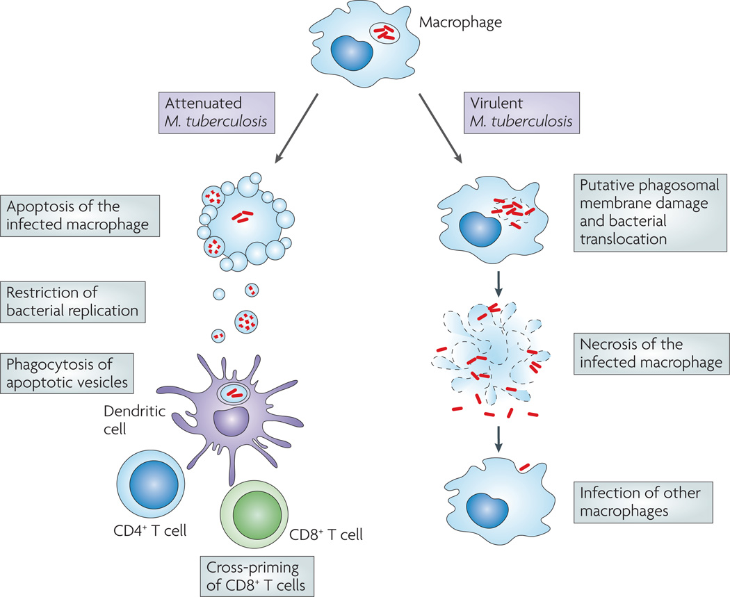
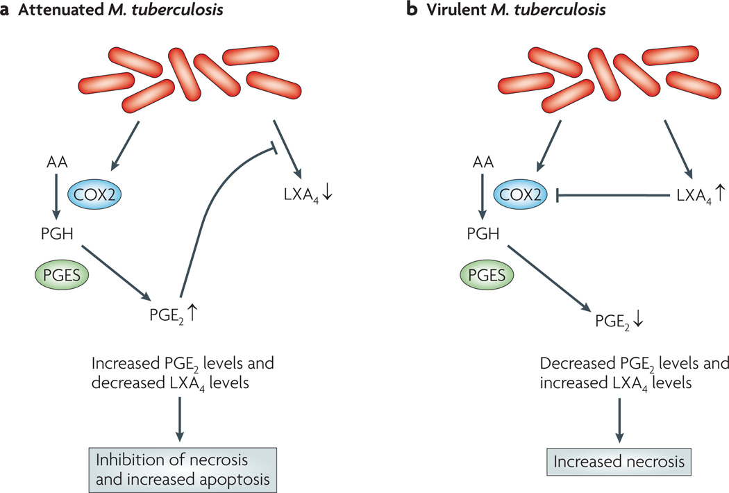
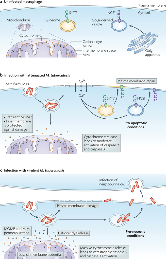
Virulent Mycobacterium tuberculosis inhibits apoptosis and triggers necrosis of host macrophages to evade innate immunity and delay the initiation of adaptive immunity. By contrast, attenuated M. tuberculosis induces macrophage apoptosis, an innate defence mechanism that reduces bacterial viability. In this Opinion article, we describe how virulent M. tuberculosis blocks production of the eicosanoid lipid mediator prostaglandin E(2) (PGE(2)). PGE(2) production by infected macrophages prevents mitochondrial damage and initiates plasma membrane repair, two processes that are crucial for preventing necrosis and inducing apoptosis. Thus, M. tuberculosis-mediated modulation of eicosanoid production determines the death modality of the infected macrophage, which in turn has a substantial impact on the outcome of infection.


Figures

Figure 1
Figure 1. The fate of infected macrophages affects host resistance to Mycobacterium tuberculosis infection
Mycobacterium tuberculosis infects macrophages and then survives and replicates in the phagosome. Macrophages infected with attenuated strains of M. tuberculosis undergo apoptosis, a death modality that impairs bacterial replication. Apoptosis of infected macrophages provides an important link to adaptive immunity, as apoptotic vesicles containing bacterial antigens are taken up by dendritic cells. The dendritic cells can efficiently present these antigens to naive T cells, leading to their activation. By contrast, virulent M. tuberculosis inhibits apoptosis and, instead, induces necrosis. We propose that damage to the phagosomal membrane facilitates bacterial translocation into the cytosol and is a precursor to the full-scale induction of macrophage necrosis. Necrosis leads to intercellular dissemination of M. tuberculosis, as extracellular M. tuberculosis can infect other macrophages that have been recruited to the lung.
Figure 2
Figure 2. The balance of prostaglandin E2 and lipoxin A4 determine the cellular fate of macrophages infected with Mycobacterium tuberculosis
a | Infection with attenuated Mycobacterium tuberculosis strongly induces cyclooxygenase 2 (COX2) synthesis, which leads to prostaglandin E2 (PGE2) production. PGE2 has at least three actions: signalling through PGE2 receptor 2 (EP2; also known as PTGER2) protects mitochondria from inner-membrane damage and prevents loss of mitochondrial membrane potential; it also enables the repair of plasma membrane damage, an activity that is probably mediated by EP4; and it inhibits the production of lipoxin A4 (LXA4). These actions all prevent necrosis of the infected macrophage and, instead, trigger apoptosis, an innate defence mechanism. b | Virulent M. tuberculosis strongly induces the production of LXA4, which prevents the accumulation of COX2 mRNA. Consequently, prostaglandin biosynthesis falls. Without the protective actions of PGE2, the infected macrophage is more likely to undergo necrosis, a form of cell death that allows the bacterium to evade innate immunity and T cell-mediated immunity. AA, arachidonic acid; PGES, PGE synthase (also known as PTGES). Figure is modified, with permission, from REF. © (2008) The Rockefeller University Press.
Figure 3
Figure 3. The antinecrotic action of prostaglandin E2 is mediated through the induction of membrane repair and the protection of mitochondria
a | In an uninfected macrophage, different Ca2+-regulated sensors are expressed in different vesicular compartments. Thus, the lysosome contains synaptotagmin 7 (SYT7), and the Golgi-derived vesicles contain neuronal calcium sensor 1 (NCS1). The mitochondrion has a double membrane consisting of the mitochondrial inner membrane (MIM) and mitochondrial outer membrane (MOM). The mitochondrial intermembrane space contains pro-apoptotic components, including cytochrome c. The inner leaflet of the MIM is negatively charged, which leads to specific binding of cationic dyes. b | Membrane microdisruptions develop during infection with attenuated Mycobacterium tuberculosis, and the resulting entry of Ca2+ into the cell triggers the recruitment of lysosomes and Golgi-derived vesicles, both of which act as membrane donors for the repair of membrane damage. Prostaglandin E2 (PGE2) is required for membrane repair, because synthesis of the lysosomal Ca2+ sensor SYT7, which is essential for the recruitment of lysosomes to the membrane lesions, depends on PGE2. Attenuated M. tuberculosis leads to MOM permeabilization (referred to as MOMP) but leaves the MIM intact. This allows cytochrome c to leak into the cytosol and leads to the activation of caspase 9, which contributes to the activation of caspase 3 and causes apoptosis. c | Infection with virulent M. tuberculosis induces the production of lipoxin A4, which inhibits the production of cylooxygenase 2 and effectively prevents prostaglandin biosynthesis. In the absence of PGE2, plasma membrane microdisruptions remain unrepaired. Concurrently, virulent M. tuberculosis induces MOMP and permeabilization of the MIM, which parallels the loss of mitochondrial membrane potential that occurs during necrosis. These conditions trigger macrophage necrosis. We propose that virulent M. tuberculosis also disrupts the phagosomal membrane and, by inhibiting membrane repair, facilitates bacterial translocation from the phagosome into the cytosol, which is a prerequisite for exit from the dying macrophage. Thus, bacterial inhibition of prostaglandin production is an immune evasion strategy that allows M. tuberculosis to avoid the detrimental consequences of apoptosis and to exit the macrophage and propagate the infection.

Similar articles

Cited by

References

    1. Barry CE, 3rd, et al. The spectrum of latent tuberculosis: rethinking the biology and intervention strategies. Nature Rev. Microbiol. 2009;7:845–855. - PMC - PubMed
    1. Bhatt K, Salgame P. Host innate immune response to Mycobacterium tuberculosis. J. Clin. Immunol. 2007;27:347–362. - PubMed
    1. Baena A, Porcelli SA. Evasion and subversion of antigen presentation by Mycobacterium tuberculosis. Tissue Antigens. 2009;74:189–204. - PMC - PubMed
    1. Fink SL, Bergsbaken T, Cookson BT. Anthrax lethal toxin and Salmonella elicit the common cell death pathway of caspase-1-dependent pyroptosis via distinct mechanisms. Proc. Natl Acad. Sci. USA. 2008;105:4312–4317. - PMC - PubMed
    1. Kroemer G, et al. Classification of cell death: recommendations of the nomenclature committee on cell death 2009. Cell Death Differ. 2009;16:3–11. - PMC - PubMed

Publication types