. 2009 Aug;22(3):147-55.
doi: 10.1055/s-0029-1236158.

Imaging update: acute colonic diverticulitis

Affiliations

Imaging update: acute colonic diverticulitis

Kristen K Destigter et al. Clin Colon Rectal Surg. 2009 Aug.

Abstract

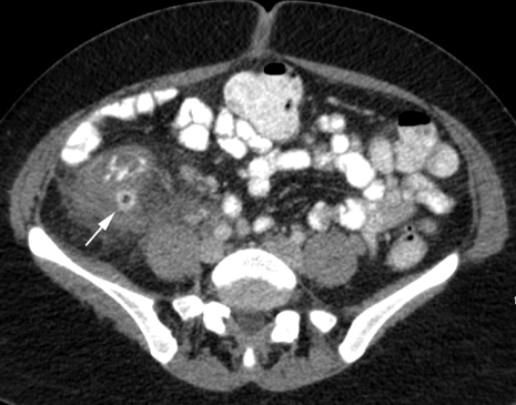
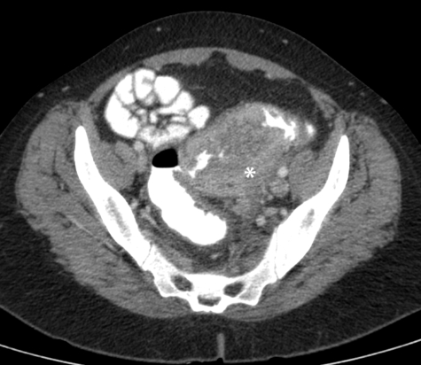
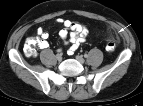
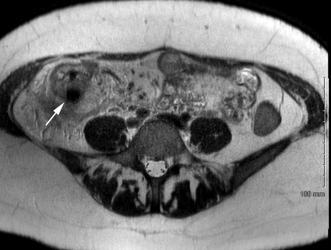
Because the incidence of colonic diverticulosis is high in the general population, incidental asymptomatic diverticulosis is commonly seen on radiology imaging studies. However, diagnostic imaging performed specifically for diverticular disease is essentially limited to imaging of suspected acute colonic diverticulitis (ACD) and its complications. The clinical diagnosis of ACD can be challenging, and imaging has become an essential tool to aid in diagnosis, assess severity of disease, and aid in treatment planning. Computed tomography (CT) has replaced contrast enema as the imaging procedure of choice for diverticulitis. Ultrasound has also been successfully used for diagnosis, and magnetic resonance imaging (MRI) has significant potential as a radiation-free imaging test for acute colonic diverticulitis.

Keywords: Acute colonic diverticulitis; computed tomography; contrast enema; diagnostic imaging; magnetic resonance imaging; ultrasound.


Figures

Figure 1
Figure 1
Diverticulitis. Axial computed tomography (CT) image of a 27-year-old man showing sigmoid wall thickening (arrow) and adjacent fat stranding (arrowhead).
Figure 2
Figure 2
Diverticulosis. Axial computed tomography (CT) image in a 49-year-old man with diverticulosis. Rectally administered contrast helps define thickened sigmoid wall, staggered haustra, and diverticular outpouchings. Note absence of adjacent fat stranding.
Figure 3
Figure 3
Mild diverticulitis. Computed tomography (CT) images from a 54-year-old woman with fever, vomiting, and left lower quadrant pain. (A) Mild sigmoid wall thickening and fat stranding around an indistinct diverticulum (arrow). (B) Image in coronal plane shows the inflamed diverticulum (arrow) at the superior aspect of mid sigmoid (U denotes uterine fundus).
Figure 4
Figure 4
Severe diverticulitis with perforation. Computed tomography (CT) image of a 44-year-old man presenting with abdominal pain, fever, and rebound tenderness. Axial image shows sigmoid wall thickening (asterisk) and extensive fat stranding as well as small amounts of free fluid, extraluminal air (arrowhead), and trace extravasated luminal contrast (arrow).
Figure 5
Figure 5
Hepatic abscess. Axial computed tomography (CT) of 56-year-old man with sigmoid diverticulitis with multiloculated fluid collection (asterisk) in left lobe of liver, subsequently drained under CT guidance.
Figure 6
Figure 6
Colovesicular fistula secondary to diverticulitis. Axial computed tomography (CT) images of an 82-year-old woman with fever and chills despite antibiotics for urinary tract infection. (A) Inflamed sigmoid with trace of air in fistula tract (arrowhead) extending to urinary bladder. (B) Ventral air (asterisk) and dependent traces of rectally administered contrast (arrow) within the urinary bladder.
Figure 7
Figure 7
Transgluteal percutaneous drainage of diverticular abscess. (A) Axial computed tomography (CT) image of 64-year-old man with fever and 3.5 cm rim enhancing fluid collection (asterisk) containing foci of air adjacent to sigmoid. (B) Image taken at conclusion of CT-guided percutaneous drain placement via transgluteal approach. (C) Fluoroscopic image from abscessogram at 1 week shows decompression of abscess cavity, but fistulous communication to rectosigmoid, outlined by contrast injected through a tube (asterisks). (D) Repeat tube injection shows decompressed abscess and lack of filling of rectosigmoid indicating resolution.
Figure 8
Figure 8
Anterior percutaneous drainage of diverticular abscess. (A) Axial computed tomography (CT) of 55-year-old woman with large diverticular abscess in cul de sac (asterisk). Note foci of air within endometrial cavity (arrowhead). (B) Computed tomography (CT) image obtained at conclusion of percutaneous drainage procedure showing pigtail catheter in abscess cavity. (C) Oblique radiograph from abscessogram 10 days after procedure shows contrast injected through drain filling the residual abscess cavity (asterisk) with fistulous communication to right fallopian tube (arrow), endometrial cavity (arrowhead) and vagina (v). (D) Eventual follow-up CT after drain removal shows normal uterus (U) and resolved abscess.
Figure 9
Figure 9
Right-sided diverticulitis. Computed tomography (CT) image of a 30-year-old woman with right abdominal pain obtained after ultrasound showed a normal gallbladder. CT shows ascending colonic wall thickening with fat stranding around inflamed diverticulum (arrow).
Figure 10
Figure 10
Adenocarcinoma. Axial computed tomography (CT) image of a 42-year-old woman presenting with weight loss and lower abdominal pain shows marked wall thickening of sigmoid (asterisk) with left pelvic sidewall involvement. Note lack of fat stranding anterior to sigmoid.
Figure 11
Figure 11
Epiploic appendagitis. Axial computed tomography (CT) image of a 42-year-old man with acute left lower quadrant pain caused by epiploic appendagitis, with ovoid fat attenuation structure (arrow) with adjacent fat stranding, anterior to the normal proximal sigmoid colon.
Figure 12
Figure 12
Magnetic resonance image (MRI) of right-sided diverticulitis. Axial MRI of a 30-year-old woman with right abdominal pain obtained after ultrasound showed a normal gallbladder (same patient as Fig. 9). T2-weighted MRI at same level as CT shows ascending colonic wall thickening with fat stranding around inflamed diverticulum (arrow).

Similar articles

Cited by

References

    1. Baker M E. Imaging and interventional techniques in acute left-sided diverticulitis. J Gastrointest Surg. 2008;12(8):1314–1317. - PubMed
    1. Balthazar E J, Megibow A, Schinella R A, Gordon R. Limitations in the CT diagnosis of acute diverticulitis: comparison of CT, contrast enema, and pathologic findings in 16 patients. AJR Am J Roentgenol. 1990;154(2):281–285. - PubMed
    1. Sheiman L, Levine M S, Levin A A, et al. Chronic diverticulitis: clinical, radiographic, and pathologic findings. AJR Am J Roentgenol. 2008;191(2):522–528. - PubMed
    1. Hulnick D H, Megibow A J, Balthazar E J, Gordon R B, Surapenini R, Bosniak M A. Perforated colorectal neoplasms: correlation of clinical, contrast enema, and CT examinations. Radiology. 1987;164(3):611–615. - PubMed
    1. Rao P M, Rhea J T, Novelline R A, et al. Helical CT with only colonic contrast material for diagnosing diverticulitis: prospective evaluation of 150 patients. AJR Am J Roentgenol. 1998;170(6):1445–1449. - PubMed