

Toward reconstructing spike trains from large-scale calcium imaging data
- PMID: 20676302
- PMCID: PMC2880024
- DOI: 10.2976/1.3284977
Toward reconstructing spike trains from large-scale calcium imaging data
Abstract
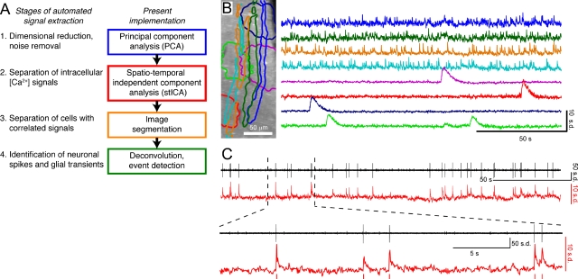
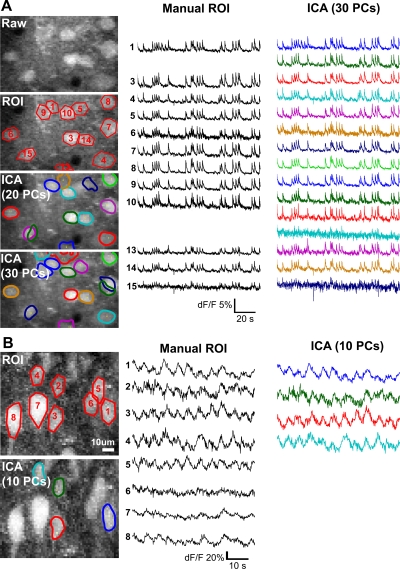
Neural activity can be captured by state-of-the-art optical imaging methods although the analysis of the resulting data sets is often manual and not standardized. Therefore, laboratories using large-scale calcium imaging eagerly await software toolboxes that can automate the process of identifying cells and inferring spikes. An algorithm proposed and implemented in a recent paper by Mukamel et al. [Neuron 63, 747-760 (2009)] used independent component analysis and offers significant improvements over conventional methods. The approach should be widely applicable, as tested with data obtained from the mouse cerebellum, neocortex, and spinal cord. The emergence of analysis tools in parallel with the rapid advances in optical imaging is an exciting development that will stimulate new discoveries and further elucidate the functions of neural circuits.
Figures
Comment on
-
Automated analysis of cellular signals from large-scale calcium imaging data.Neuron. 2009 Sep 24;63(6):747-60. doi: 10.1016/j.neuron.2009.08.009. Neuron. 2009. PMID: 19778505 Free PMC article.
Similar articles
-
NeuroCa: integrated framework for systematic analysis of spatiotemporal neuronal activity patterns from large-scale optical recording data.Neurophotonics. 2015 Jul;2(3):035003. doi: 10.1117/1.NPh.2.3.035003. Epub 2015 Jul 28. Neurophotonics. 2015. PMID: 26229973 Free PMC article.
-
Synaptic transmission of chaotic spike trains between primary afferent fiber and spinal dorsal horn neuron in the rat.Neuroscience. 2004;125(4):1051-60. doi: 10.1016/j.neuroscience.2004.02.035. Neuroscience. 2004. PMID: 15120864
-
Wavelet-based processing of neuronal spike trains prior to discriminant analysis.J Neurosci Methods. 2004 Apr 30;134(2):159-68. doi: 10.1016/j.jneumeth.2003.11.007. J Neurosci Methods. 2004. PMID: 15003382
-
Dendritic Spikes in Sensory Perception.Front Cell Neurosci. 2017 Feb 15;11:29. doi: 10.3389/fncel.2017.00029. eCollection 2017. Front Cell Neurosci. 2017. PMID: 28261060 Free PMC article. Review.
-
Emerging ideas and tools to study the emergent properties of the cortical neural circuits for voluntary motor control in non-human primates.F1000Res. 2019 May 29;8:F1000 Faculty Rev-749. doi: 10.12688/f1000research.17161.1. eCollection 2019. F1000Res. 2019. PMID: 31275561 Free PMC article. Review.
Cited by
-
Spatiotemporal dynamics of rhythmic spinal interneurons measured with two-photon calcium imaging and coherence analysis.J Neurophysiol. 2010 Dec;104(6):3323-33. doi: 10.1152/jn.00679.2010. Epub 2010 Sep 22. J Neurophysiol. 2010. PMID: 20861442 Free PMC article.
References
-
- Busche, M A, Eichhoff, G, Adelsberger, H, Abramowski, D, Wiederhold, K H, Haass, C, Staufenbiel, M, Konnerth, A, and Garaschuk, O (2008). “Clusters of hyperactive neurons near amyloid plaques in a mouse model of Alzheimer's disease.” Science SCIEAS 321, 1686–1689.10.1126/science.1162844 - DOI - PubMed
LinkOut - more resources
Full Text Sources