. 2010 Jul 22;6(7):e1000861.
doi: 10.1371/journal.pcbi.1000861.

G-quadruplex DNA sequences are evolutionarily conserved and associated with distinct genomic features in Saccharomyces cerevisiae

Affiliations

G-quadruplex DNA sequences are evolutionarily conserved and associated with distinct genomic features in Saccharomyces cerevisiae

John A Capra et al. PLoS Comput Biol. .

Abstract

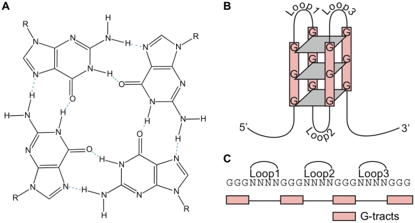
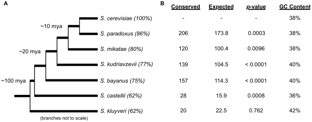
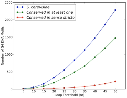
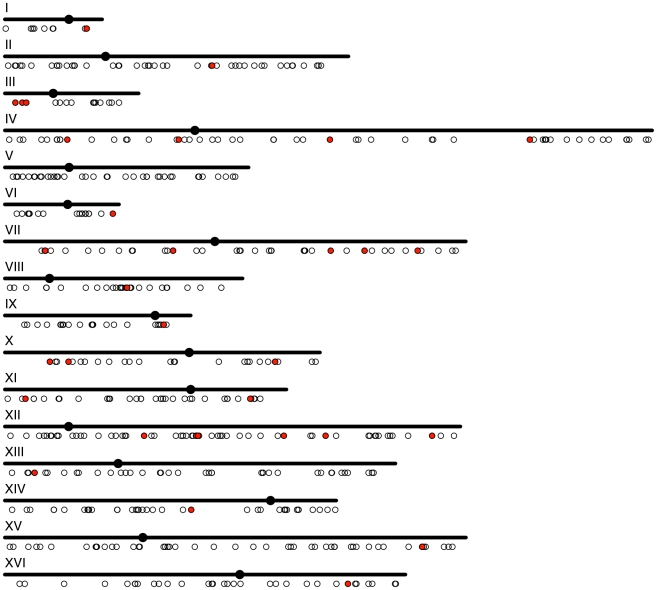
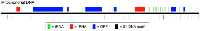
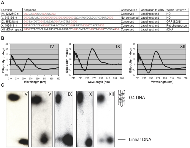
G-quadruplex DNA is a four-stranded DNA structure formed by non-Watson-Crick base pairing between stacked sets of four guanines. Many possible functions have been proposed for this structure, but its in vivo role in the cell is still largely unresolved. We carried out a genome-wide survey of the evolutionary conservation of regions with the potential to form G-quadruplex DNA structures (G4 DNA motifs) across seven yeast species. We found that G4 DNA motifs were significantly more conserved than expected by chance, and the nucleotide-level conservation patterns suggested that the motif conservation was the result of the formation of G4 DNA structures. We characterized the association of conserved and non-conserved G4 DNA motifs in Saccharomyces cerevisiae with more than 40 known genome features and gene classes. Our comprehensive, integrated evolutionary and functional analysis confirmed the previously observed associations of G4 DNA motifs with promoter regions and the rDNA, and it identified several previously unrecognized associations of G4 DNA motifs with genomic features, such as mitotic and meiotic double-strand break sites (DSBs). Conserved G4 DNA motifs maintained strong associations with promoters and the rDNA, but not with DSBs. We also performed the first analysis of G4 DNA motifs in the mitochondria, and surprisingly found a tenfold higher concentration of the motifs in the AT-rich yeast mitochondrial DNA than in nuclear DNA. The evolutionary conservation of the G4 DNA motif and its association with specific genome features supports the hypothesis that G4 DNA has in vivo functions that are under evolutionary constraint.


Conflict of interest statement

The authors have declared that no competing interests exist.

Figures

Figure 1
Figure 1. The G4 DNA structure and motif.
(A) Structure of a G-quartet. The planar ring of four hydrogen-bonded guanines is formed by guanines from different G-tracts, which are separated by intervening loop regions in the intra-molecular G4 DNA structure. (B) Schematic of an intra-molecular G4 DNA structure consisting of three G-quartets. Inter-molecular G4 DNA structures can also form from two or four strands. (C) The G4 DNA motif sequence used in this study with four G-tracts of three guanines separated by loop regions.
Figure 2
Figure 2. The evolutionary conservation of G4 DNA motifs between S. cerevisiae and six related yeast species.
(A) The phylogenetic tree for the seven yeast species considered in this study (not to scale). The four sensu stricto species (S. paradoxus, S. mikatae, S. kudriavzevii, S. bayanus) diverged from S. cerevisiae within the last ∼20 million years. S. castelli and S. kluyveri are considerably more distant . The percent sequence identity to S. cerevisiae over the alignable regions is given in parentheses. (B) The evolutionary conservation of the 507 non-telomeric, nuclear S. cerevisiae G4 DNA motifs in sequence regions that could be aligned to at least one other genome. Significantly more G4 DNA motifs were conserved than expected by chance between S. cerevisiae and five of the six species considered. The one exception was S. kluyveri, which is the most distant and GC-rich species among the seven yeasts.
Figure 3
Figure 3. The conservation of G4 DNA motifs as a function of the loop length threshold.
The number of G4 DNA motifs identified in S. cerevisiae increases as maximum loop length limit is increased (blue line). More than half of the motifs are conserved in at least one other species at each loop threshold (green line). The number of motifs conserved in all sensu stricto species also increases as longer loops are tolerated (red line).
Figure 4
Figure 4. The distribution of G4 DNA motifs across the S. cerevisiae nuclear genome.
Each small circle represents the location of a G4 DNA motif in a chromosome. Motifs conserved across the sensu stricto species are highlighted in red.
Figure 5
Figure 5. The distribution of G4 DNA motifs across the S. cerevisiae mitochondrial DNA.
The horizontal black line represents the 75kb mtDNA genome. The rectangles above mark the location of tRNA genes (green), rRNA genes (red), and ORFs (blue). ORFs that encode multiple genes (like COX1, subunit I of cytochrome c oxidase) are drawn as a single rectangle. The vertical black lines below indicate the location of the 32 G4 DNA motifs across the mtDNA. The width of these lines reflects the actual length of the G4 DNA motif. (Note that several motifs are so close that they overlap in the figure.) All motifs are drawn below the mtDNA sequence regardless of the strand on which they occur. The distribution of mitochondiral G4 DNA motifs is biased against overlapping these genomic features; only two of the 32 motifs overlap a tRNA, rRNA, or ORF (q<0.001).
Figure 6
Figure 6. G4 DNA motifs form G4 DNA structures in vitro.
(A) Characterization of the five experimentally tested G4 DNA motifs. The motifs were selected to represent a range of genome locations and conditions. (B) Circular dichroism analysis of the three of the five G4 DNA motifs tested demonstrates that they form parallel G4 DNA structures. The G4 motif tested is indicated by the chromosome number above the graph. (C) Native acrylamide gel comparing the migration of oligonucleotides before and after the formation of the G4 DNA structure. The G4 DNA structure (indicated by the cartoon on the right) migrates more slowly than linear DNA. The G4 motif tested is indicated by the chromosome number beneath the gel.

Similar articles

Cited by

References

    1. Sen D, Gilbert W. Formation of parallel four-stranded complexes by guanine-rich motifs in DNA and its implications for meiosis. Nature. 1988;334:364–366. - PubMed
    1. Sundquist WI, Klug A. Telomeric DNA dimerizes by formation of guanine tetrads between hairpin loops. Nature. 1989;342:825–829. - PubMed
    1. Burge S, Parkinson GN, Hazel P, Todd AK, Neidle S. Quadruplex DNA: sequence, topology and structure. Nucleic Acids Res. 2006;34:5402–5415. - PMC - PubMed
    1. Stegle O, Payet L, Mergny JL, MacKay DJ, Leon JH. Predicting and understanding the stability of G-quadruplexes. Bioinformatics. 2009;25:i374–382. - PMC - PubMed
    1. Maizels N. Dynamic roles for G4 DNA in the biology of eukaryotic cells. Nat Struct Mol Biol. 2006;13:1055–1059. - PubMed

Publication types