

Extracellular and mixotrophic symbiosis in the whale-fall mussel Adipicola pacifica: a trend in evolution from extra- to intracellular symbiosis
- PMID: 20676405
- PMCID: PMC2910738
- DOI: 10.1371/journal.pone.0011808
Extracellular and mixotrophic symbiosis in the whale-fall mussel Adipicola pacifica: a trend in evolution from extra- to intracellular symbiosis
Abstract
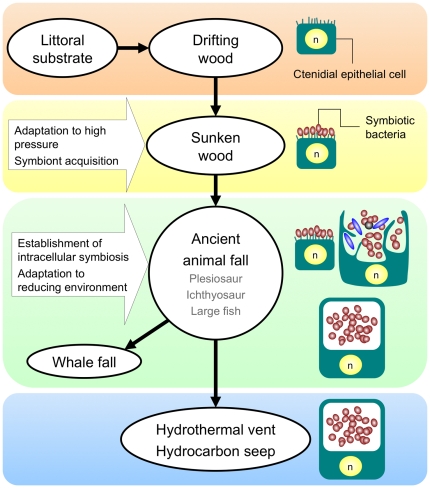
Background: Deep-sea mussels harboring chemoautotrophic symbionts from hydrothermal vents and seeps are assumed to have evolved from shallow-water asymbiotic relatives by way of biogenic reducing environments such as sunken wood and whale falls. Such symbiotic associations have been well characterized in mussels collected from vents, seeps and sunken wood but in only a few from whale falls.
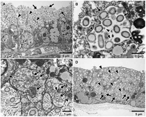
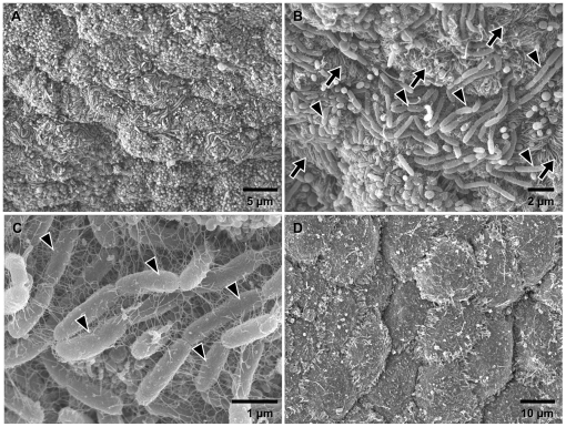
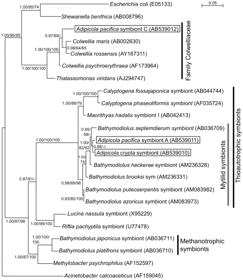
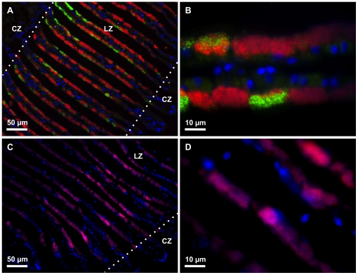
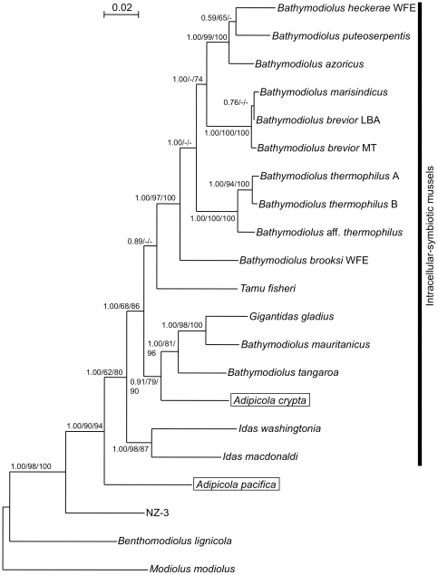
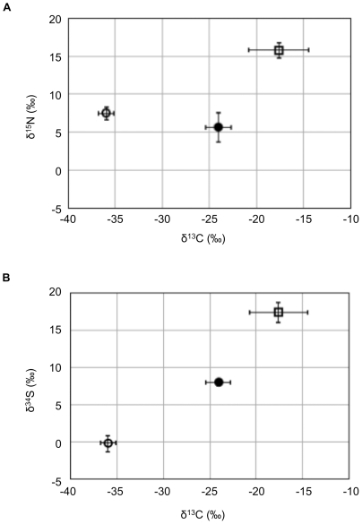
Methodology/principal finding: Here we report symbioses in the gill tissues of two mussels, Adipicola crypta and Adipicola pacifica, collected from whale-falls on the continental shelf in the northwestern Pacific. The molecular, morphological and stable isotopic characteristics of bacterial symbionts were analyzed. A single phylotype of thioautotrophic bacteria was found in A. crypta gill tissue and two distinct phylotypes of bacteria (referred to as Symbiont A and Symbiont C) in A. pacifica. Symbiont A and the A. crypta symbiont were affiliated with thioautotrophic symbionts of bathymodiolin mussels from deep-sea reducing environments, while Symbiont C was closely related to free-living heterotrophic bacteria. The symbionts in A. crypta were intracellular within epithelial cells of the apical region of the gills and were extracellular in A. pacifica. No spatial partitioning was observed between the two phylotypes in A. pacifica in fluorescence in situ hybridization experiments. Stable isotopic analyses of carbon and sulfur indicated the chemoautotrophic nature of A. crypta and mixotrophic nature of A. pacifica. Molecular phylogenetic analyses of the host mussels showed that A. crypta constituted a monophyletic clade with other intracellular symbiotic (endosymbiotic) mussels and that A. pacifica was the sister group of all endosymbiotic mussels.
Conclusions/significance: These results strongly suggest that the symbiosis in A. pacifica is at an earlier stage in evolution than other endosymbiotic mussels. Whale falls and other modern biogenic reducing environments may act as refugia for primal chemoautotrophic symbioses between eukaryotes and prokaryotes since the extinction of ancient large marine vertebrates.
Conflict of interest statement
Figures
Similar articles
-
Dual symbiosis in a Bathymodiolus sp. mussel from a methane seep on the Gabon continental margin (Southeast Atlantic): 16S rRNA phylogeny and distribution of the symbionts in gills.Appl Environ Microbiol. 2005 Apr;71(4):1694-700. doi: 10.1128/AEM.71.4.1694-1700.2005. Appl Environ Microbiol. 2005. PMID: 15811991 Free PMC article.
-
Sulphur-oxidizing extracellular bacteria in the gills of Mytilidae associated with wood falls.FEMS Microbiol Ecol. 2008 Mar;63(3):338-49. doi: 10.1111/j.1574-6941.2008.00438.x. Epub 2008 Jan 19. FEMS Microbiol Ecol. 2008. PMID: 18218025
-
Geographical structure of endosymbiotic bacteria hosted by Bathymodiolus mussels at eastern Pacific hydrothermal vents.BMC Evol Biol. 2017 May 30;17(1):121. doi: 10.1186/s12862-017-0966-3. BMC Evol Biol. 2017. PMID: 28558648 Free PMC article.
-
Symbioses between deep-sea mussels (Mytilidae: Bathymodiolinae) and chemosynthetic bacteria: diversity, function and evolution.C R Biol. 2009 Feb-Mar;332(2-3):298-310. doi: 10.1016/j.crvi.2008.08.003. Epub 2008 Nov 25. C R Biol. 2009. PMID: 19281960 Review.
-
Symbiotic diversity in marine animals: the art of harnessing chemosynthesis.Nat Rev Microbiol. 2008 Oct;6(10):725-40. doi: 10.1038/nrmicro1992. Nat Rev Microbiol. 2008. PMID: 18794911 Review.
Cited by
-
Metatranscriptomic analysis of sulfur oxidation genes in the endosymbiont of solemya velum.Front Microbiol. 2011 Jun 20;2:134. doi: 10.3389/fmicb.2011.00134. eCollection 2011. Front Microbiol. 2011. PMID: 21738524 Free PMC article.
-
Integrative biology of Idas iwaotakii (Habe, 1958), a 'model species' associated with sunken organic substrates.PLoS One. 2013 Jul 24;8(7):e69680. doi: 10.1371/journal.pone.0069680. Print 2013. PLoS One. 2013. PMID: 23894520 Free PMC article.
-
Molecular characterization of Bathymodiolus mussels and gill symbionts associated with chemosynthetic habitats from the U.S. Atlantic margin.PLoS One. 2019 Mar 14;14(3):e0211616. doi: 10.1371/journal.pone.0211616. eCollection 2019. PLoS One. 2019. PMID: 30870419 Free PMC article.
-
Bioenergetic evolution in proteobacteria and mitochondria.Genome Biol Evol. 2014 Nov 27;6(12):3238-51. doi: 10.1093/gbe/evu257. Genome Biol Evol. 2014. PMID: 25432941 Free PMC article.
-
Identification of Associations between Bacterioplankton and Photosynthetic Picoeukaryotes in Coastal Waters.Front Microbiol. 2016 Mar 22;7:339. doi: 10.3389/fmicb.2016.00339. eCollection 2016. Front Microbiol. 2016. PMID: 27148165 Free PMC article.
References
-
- Felbeck H, Childress JJ, Somero GN. Calvin-Benson Cycle and sulphide oxidation enzymes in animals from sulphide-rich habitats. Nature. 1981;293:291–293.
-
- Corliss JB, Dymond J, Gordon LI, Edmond JM, Herzen RPv, et al. Submarine thermal springs on the Galapagos Rift. Science. 1979;203:1073–1083. - PubMed
-
- Fiala-Médioni A, Le Pennec M. Trophic structural adaptations in relation to the bacterial association of bivalve molluscs from hydrothermal vents and subduction zones. Symbiosis. 1987;4:63–74.
-
- Childress JJ, Fisher CR, Brooks JM, Kennicutt MC, II, Bidigare R, et al. A methanotrophic marine molluscan (Bivalvia, Mytilidae) symbiosis: mussels fueled by gas. Science. 1986;233:1306–1308. - PubMed
Publication types
MeSH terms
Substances
Associated data
- Actions
- Actions
- Actions
- Actions
- Actions
- Actions
- Actions
- Actions
- Actions
LinkOut - more resources
Full Text Sources
Molecular Biology Databases
