

Correlations between radiographic, magnetic resonance and histological examinations on the degeneration of human lumbar intervertebral discs
- PMID: 20676571
- PMCID: PMC10938976
- DOI: 10.1590/s1516-31802010000200004
Correlations between radiographic, magnetic resonance and histological examinations on the degeneration of human lumbar intervertebral discs
Abstract
Context and objective: There is controversy regarding which imaging method is best for identifying early degenerative alterations in intervertebral discs. No correlations between such methods and histological finds are presented in the literature. The aim of this study was to correlate the thickness of intervertebral discs measured on simple radiographs with the degree of degeneration seen on magnetic resonance images and the histological findings relating to nerve ends inside the discs.
Design and setting: Cross-sectional correlation study on the lumbar spines of human cadavers, at Universidade Federal de São Paulo (Unifesp), São Paulo, Brazil.
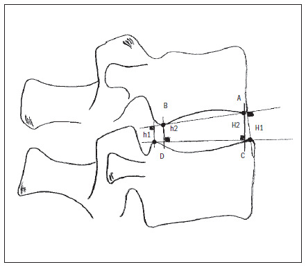
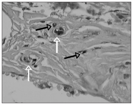
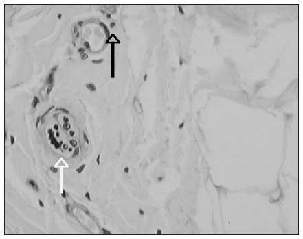
Methods: Ten lumbar spinal columns were extracted from human cadavers and subjected to magnetic resonance imaging and simple radiography. They were classified according to the degree of disc degeneration seen on magnetic resonance, and the thickness of the discs was measured on radiographs. The intervertebral discs were then extracted, embedded in paraffin and analyzed immunohistochemically with protein S100, and the nerve fibers were counted and classified.
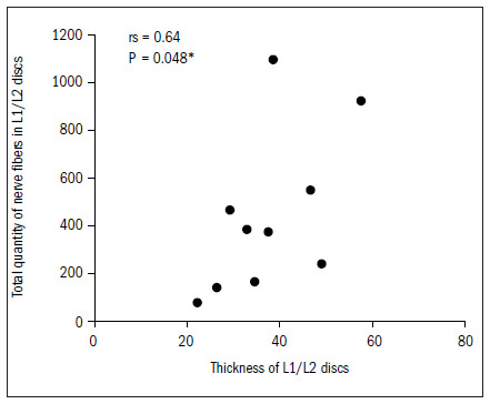
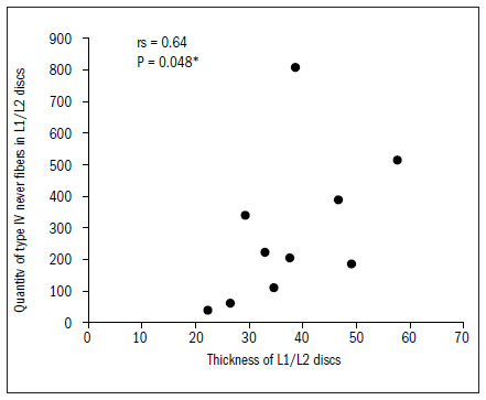
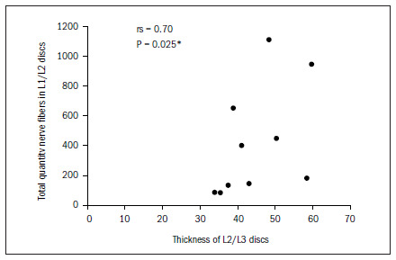
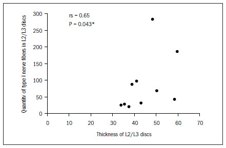
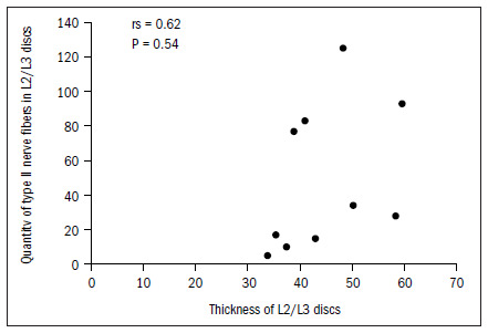
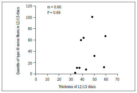
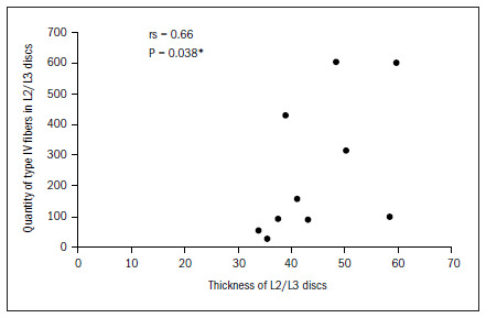
Results: No correlation was observed between the thickness of the intervertebral discs and the degree of degeneration seen on magnetic resonance images. Only the uppermost lumbar discs (L1/L2 and L2/L3) presented a correlation between their thickness and type I and IV nerve endings.
Conclusion: Reduced disc thickness is unrelated to increased presence of nerve ends in intervertebral discs, or to the degree of disc degeneration.
Contexto E Objetivo:: Há controvérsia sobre qual o melhor método de imagem para identificar alterações degenerativas precoces do disco intervertebral. Falta na literatura correlação desses métodos com os achados histológicos. O objetivo deste estudo foi relacionar a altura dos discos intervertebrais medidos em radiografias simples com o grau de degeneração nas imagens de ressonância magnética e os achados histológicos das terminações nervosas encontradas no interior do disco.
Tipo De Estudo E Local:: Estudo transversal de correlação em coluna lombar de cadáveres humanos, na Universidade Federal de São Paulo (Unifesp), São Paulo, Brasil.
Métodos:: Dez colunas lombares foram retiradas de cadáveres humanos e submetidas a imagens de ressonância magnética e radiografias simples. Foram classificadas de acordo com o grau de degeneração dos discos pela ressonância e mensuradas as alturas dos discos nas radiografias. Os discos intervertebrais foram retirados, incluídos em parafina e foi realizado estudo imunoistoquímico com proteína S100; as fibras nervosas foram contadas e classificadas.
Resultados:: Não foi observada correlação entre a altura dos discos intervertebrais com o grau de degeneração nas imagens de ressonância magnética. Apenas os discos lombares altos (L1/L2 e L2/L3) apresentaram correlação entre a altura e as terminações nervosas dos tipos I e IV.
Conclusão:: A diminuição da altura dos discos não está relacionada ao aumento de terminações nervosas nos discos intervertebrais e nem com o grau de degeneração dos discos.
Figures
References
-
- Andersson GB. Epidemiology of low back pain. Acta Orthop Scand Suppl. 1998;281:28–31. - PubMed
-
- Battié MC, Videman T, Parent E. Lumbar disc degeneration: epidemiology and genetic influences. Spine (Phila Pa 1976) 2004;29(23):2679–2690. - PubMed
-
- Peterson CK, Bolton JE, Wood AR. A cross-sectional study correlating lumbar spine degeneration with disability and pain. Spine (Phila Pa 1976) 2000;25(2):218–223. - PubMed
-
- Luoma K, Riihimäki H, Luukkonen R, Raininko R, Viikari-Juntura E, Lamminen A. Low back pain in relation to lumbar disc degeneration. Spine (Phila Pa 1976) 2000;25(4):487–492. - PubMed
Publication types
MeSH terms
Substances
LinkOut - more resources
Full Text Sources
