

In vitro quantification of wear in tibial inserts using microcomputed tomography
- PMID: 20676810
- PMCID: PMC3008888
- DOI: 10.1007/s11999-010-1490-6
In vitro quantification of wear in tibial inserts using microcomputed tomography
Abstract
Background: Wear of polyethylene tibial inserts can decrease the longevity of total knee arthroplasty. Wear is currently assessed using laboratory methods that may not permit backside wear measurements or do not quantify surface deviation.
Questions/purposes: We developed and validated a technique to quantify polyethylene wear in tibial inserts using microcomputed tomography (micro-CT), a nondestructive high-resolution imaging technique that provides detailed images of surface geometry in addition to volumetric measurements.
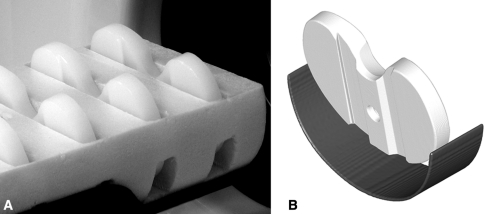
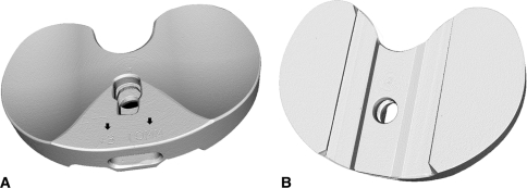
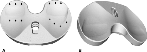
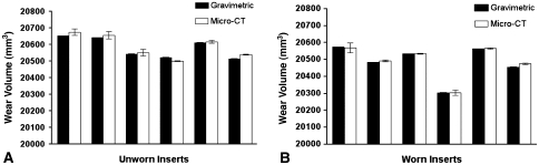
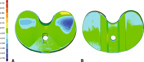
Methods: Six unworn and six wear-simulated polyethylene tibial inserts were evaluated. Each insert was scanned three times using micro-CT at a resolution of 50 μm. The insert surface was reconstructed for each scan and the insert volume was calculated. Gravimetric analysis was performed for all inserts, and the micro-CT and gravimetric volumes were compared to determine accuracy. We created three-dimensional surface deviation maps.
Results: Micro-CT generated high-quality three-dimensional renderings of the insert surface geometry. Between-scan precision was 0.07%; we observed no difference between micro-CT and gravimetric volume measurements.
Conclusions: Micro-CT can provide precise and accurate volumetric measurements in addition to quantifiable three-dimensional surface deviation maps for the entire insert surface. The technique has the potential to evaluate wear in wear simulator trials and retrieval studies.
Clinical relevance: This micro-CT technique combines the benefits of volumetric and surface scanning methods to quantify wear across all surfaces of polyethylene components with a single tool. When applied in wear simulator and retrieval studies, these measurements can be used to evaluate and predict the wear properties of the components.
Figures
Similar articles
-
Can microcomputed tomography measure retrieved polyethylene wear? Comparing fixed-bearing and rotating-platform knees.Clin Orthop Relat Res. 2013 Jan;471(1):86-93. doi: 10.1007/s11999-012-2513-2. Clin Orthop Relat Res. 2013. PMID: 22879092 Free PMC article.
-
How do CAD models compare with reverse engineered manufactured components for use in wear analysis?Clin Orthop Relat Res. 2012 Jul;470(7):1847-54. doi: 10.1007/s11999-011-2143-0. Clin Orthop Relat Res. 2012. PMID: 22016002 Free PMC article.
-
Does increased topside conformity in modular total knee arthroplasty lead to increased backside wear?Clin Orthop Relat Res. 2015 Jan;473(1):220-5. doi: 10.1007/s11999-014-3648-0. Clin Orthop Relat Res. 2015. PMID: 24777725 Free PMC article.
-
All-polyethylene tibial components in modern total knee arthroplasty.J Am Acad Orthop Surg. 2011 Sep;19(9):527-35. doi: 10.5435/00124635-201109000-00003. J Am Acad Orthop Surg. 2011. PMID: 21885698 Review.
-
Volumetric Tissue Imaging of Surgical Tissue Specimens Using Micro-Computed Tomography: An Emerging Digital Pathology Modality for Nondestructive, Slide-Free Microscopy-Clinical Applications of Digital Pathology in 3 Dimensions.Am J Clin Pathol. 2023 Mar 13;159(3):242-254. doi: 10.1093/ajcp/aqac143. Am J Clin Pathol. 2023. PMID: 36478204
Cited by
-
Micro X-Ray Computed Tomography Mass Loss Assessment of Different UHMWPE: A Hip Joint Simulator Study on Standard vs. Cross-Linked Polyethylene.PLoS One. 2017 Jan 20;12(1):e0170263. doi: 10.1371/journal.pone.0170263. eCollection 2017. PLoS One. 2017. PMID: 28107468 Free PMC article.
-
Relationship of surface damage appearance and volumetric wear in retrieved TKR polyethylene liners.J Biomed Mater Res B Appl Biomater. 2017 Oct;105(7):2053-2059. doi: 10.1002/jbm.b.33684. Epub 2016 Jul 12. J Biomed Mater Res B Appl Biomater. 2017. PMID: 27401236 Free PMC article.
-
Wear Distribution Detection of Knee Joint Prostheses by Means of 3D Optical Scanners.Materials (Basel). 2017 Mar 30;10(4):364. doi: 10.3390/ma10040364. Materials (Basel). 2017. PMID: 28772725 Free PMC article.
-
Current Total Knee Designs: Does Baseplate Roughness or Locking Mechanism Design Affect Polyethylene Backside Wear?Clin Orthop Relat Res. 2017 Dec;475(12):2970-2980. doi: 10.1007/s11999-017-5494-3. Epub 2017 Sep 13. Clin Orthop Relat Res. 2017. PMID: 28905208 Free PMC article.
-
Can microcomputed tomography measure retrieved polyethylene wear? Comparing fixed-bearing and rotating-platform knees.Clin Orthop Relat Res. 2013 Jan;471(1):86-93. doi: 10.1007/s11999-012-2513-2. Clin Orthop Relat Res. 2013. PMID: 22879092 Free PMC article.
References
-
- Blunt L, Bills P, Jiang X, Chakrabarty G. Improvement in the assessment of wear of total knee replacements using coordinate-measuring machine techniques. Proc Inst Mech Eng H. 2008;222:309–318. - PubMed
-
- Conditt MA, Stein JA, Noble PC. Factors affecting the severity of backside wear of modular tibial inserts. J Bone Joint Surg Am. 2004;86:305–311. - PubMed
Publication types
MeSH terms
Substances
LinkOut - more resources
Full Text Sources
Medical
