

Methotrexate-related Epstein-Barr Virus (EBV)-associated lymphoproliferative disorder--so-called "Hodgkin-like lesion"--of the oral cavity in a patient with rheumatoid arthritis
- PMID: 20676828
- PMCID: PMC2996501
- DOI: 10.1007/s12105-010-0202-6
Methotrexate-related Epstein-Barr Virus (EBV)-associated lymphoproliferative disorder--so-called "Hodgkin-like lesion"--of the oral cavity in a patient with rheumatoid arthritis
Abstract
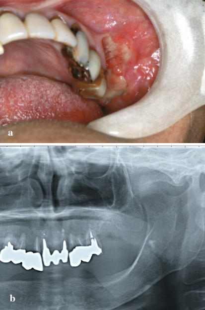
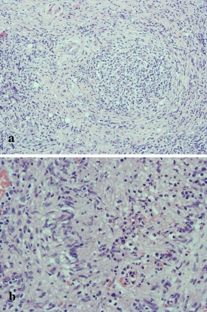
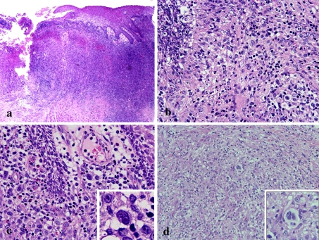
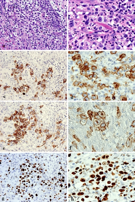
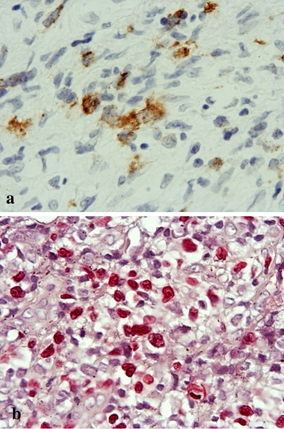
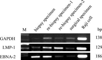
Patients affected by autoimmune diseases (rheumatoid arthritis, psoriasis, dermatomyositis) who are treated with methotrexate (MTX) sometimes develop lymphoproliferative disorders (LPDs). In approximately 40% of reported cases, the affected sites have been extranodal, and have included the gastrointestinal tract, skin, lung, kidney, and soft tissues. However, MTX-associated LPD (MTX-LPD) is extremely rare in the oral cavity. Here we report a 69-year-old Japanese woman with rheumatoid arthritis (RA) who developed MTX-LPD resembling Hodgkin's disease--so-called "Hodgkin-like lesion"--in the left upper jaw. Histopathologically, large atypical lymphoid cells including Hodgkin or Reed-Sternberg-like cells were found to have infiltrated into granulation tissue in the ulcerative oral mucosa. Immunohistochemistry showed that the large atypical cells were positive for CD20, CD30 and Epstein-Barr virus (EBV)-latent infection membrane protein-1 (LMP-1) and negative for CD15. EBV was detected by in situ hybridization (ISH) with EBV-encoded small RNA (EBER), and polymerase chain reaction (PCR) for LMP-1 and EBNA-2 in material taken from the formalin-fixed, paraffin-embedded specimen. To our knowledge, this is the first reported case of MTX-related EBV-associated LPD (MTX-EBVLPD), "Hodgkin-like lesion", of the oral cavity in a patient with RA.
Figures
Similar articles
-
Methotrexate-related Epstein-Barr virus-associated lymphoproliferative disorder occurring in the gingiva of a patient with rheumatoid arthritis.Int J Clin Exp Pathol. 2013 Sep 15;6(10):2237-41. eCollection 2013. Int J Clin Exp Pathol. 2013. PMID: 24133604 Free PMC article.
-
A case of age-related Epstein-Barr virus (EBV)-associated B cell lymphoproliferative disorder, so-called polymorphous subtype, of the mandible, with a review of the literature.Head Neck Pathol. 2013 Jun;7(2):178-87. doi: 10.1007/s12105-012-0392-1. Epub 2012 Aug 7. Head Neck Pathol. 2013. PMID: 22869357 Free PMC article. Review.
-
Plasmacytic hyperplasia in age-related Epstein-Barr virus-associated lymphoproliferative disorders: a report of two cases.Pathol Res Pract. 2008;204(4):267-72. doi: 10.1016/j.prp.2007.11.007. Epub 2008 Jan 9. Pathol Res Pract. 2008. PMID: 18187262
-
Methotrexate-associated Lymphoproliferative Disorders in Patients With Rheumatoid Arthritis: Clinicopathologic Features and Prognostic Factors.Am J Surg Pathol. 2019 Jul;43(7):869-884. doi: 10.1097/PAS.0000000000001271. Am J Surg Pathol. 2019. PMID: 31116708
-
Spontaneous regression of lymphoproliferative disorders in patients treated with methotrexate for rheumatoid arthritis and other rheumatic diseases.J Clin Oncol. 1996 Jun;14(6):1943-9. doi: 10.1200/JCO.1996.14.6.1943. J Clin Oncol. 1996. PMID: 8656264 Review.
Cited by
-
Epstein-Barr virus mucocutaneous ulcer followed by Hodgkin lymphoma in multiple myeloma patient.Clin Case Rep. 2022 Mar 3;10(3):e05528. doi: 10.1002/ccr3.5528. eCollection 2022 Mar. Clin Case Rep. 2022. PMID: 35280097 Free PMC article.
-
Overexpression of Activation-Induced Cytidine Deaminase in MTX- and Age-Related Epstein-Barr Virus-Associated B-Cell Lymphoproliferative Disorders of the Head and Neck.J Oncol. 2015;2015:605750. doi: 10.1155/2015/605750. Epub 2015 Mar 5. J Oncol. 2015. PMID: 25834572 Free PMC article.
-
Methotrexate-related Epstein-Barr virus-associated lymphoproliferative disorder occurring in the gingiva of a patient with rheumatoid arthritis.Int J Clin Exp Pathol. 2013 Sep 15;6(10):2237-41. eCollection 2013. Int J Clin Exp Pathol. 2013. PMID: 24133604 Free PMC article.
-
Recurrent Methotrexate-related Lymphoproliferative Disorder of the Lumbar Spine Origin: A Case Report.J Orthop Case Rep. 2021 Mar;11(3):63-66. doi: 10.13107/jocr.2021.v11.i03.2090. J Orthop Case Rep. 2021. PMID: 34239831 Free PMC article.
-
Clinical management for other iatrogenic immunodeficiency-associated lymphoproliferative disorders.J Clin Exp Hematop. 2019;59(2):72-92. doi: 10.3960/jslrt.19007. J Clin Exp Hematop. 2019. PMID: 31257348 Free PMC article. Review.
References
-
- Daniel M, Knowles MD. Immunodeficiency-associated lymphoproliferative disorders. Mod Pathol. 1999;12:200–217. - PubMed
-
- Salloum E, Cooper DL, Howe G, et al. Spontaneous regression of lymphoproliferative disorders in patients treated with metotrexate for rheumatoid arthritis and other rheumatic diseases. J Clin Oncol. 1996;14:1943–1949. - PubMed
Publication types
MeSH terms
Substances
LinkOut - more resources
Full Text Sources
Medical
Research Materials
